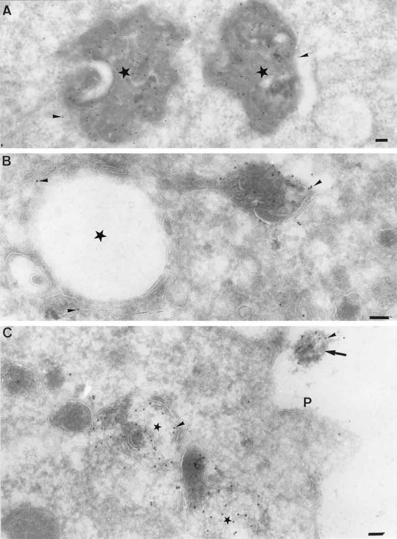
FIG. 5.
Localization of CT1a as seen by EM. MHV-infected cells were fixed at 5 (B and C) and 7 (A) h p.i., and thawed cryosections were labeled with antibodies to CT1a (arrowheads). In panel A, note extensive labeling over structures with multivesicular appearance (stars). In panel B, the CT1a-positive membranes are attached to a vacuolar structure (star), the surrounding membranes of which are also weakly labeled (arrowheads). Panel C shows another profile of CT1a-positive (arrowheads) multivesicular membranes (stars). Note the membrane structure (large arrow) at the outside of the cell that is significantly labeled with CT1a (arrowhead). P, plasma membrane. Bars, 100 nm.

