FIG. 3.
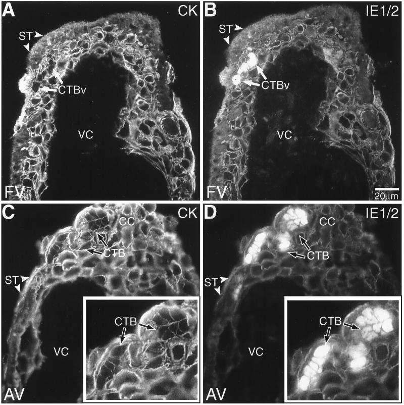
Cytotrophoblasts (CTBs) in villous explants were infected with CMV in vitro. Tissue sections prepared from both floating villi (FV; A and B) and anchoring villi (AV; C and D) were analyzed by double staining with anticytokeratin and anti-CMV IE1/2. (A) Cytokeratin (CK) staining of floating villi showed the multinucleate syncytiotrophoblasts (ST; arrowheads) that cover the villous surface and the underlying villous cytotrophoblast stem cells (CTBv; arrows). (B) CMV IE1/2 proteins were expressed by underlying clusters of infected villous cytotrophoblast stem cells. The inner stromal villous cores (VC) were consistently negative for anti-IE1/2 antibody staining. (C and D) IE1/2 protein expression was also detected in cytotrophoblasts found in the cell columns (CC) of anchoring villi. Insets show infected cytotrophoblasts at higher magnification. As a control, staining was performed as described for the experimental situation except that the primary or secondary antibody was omitted. No staining was detected (data not shown).

