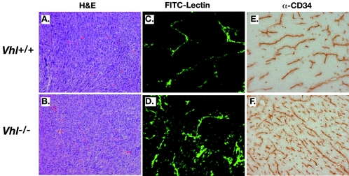
FIG. 3.
Increased vascular permeability and vessel density in Vhl−/− fibrosarcomas. (A and B) Hematoxylin and eosin staining did not indicate any significant differences in histology between Vhl+/+ and Vhl−/− fibrosarcomas. (C and D) Lectin perfusion of fibrosarcomas revealed highly organized vessel formation in Vhl+/+ tumors, while Vhl−/− tumors demonstrated increased vascular permeability as noted by an increase in background fluorescence. (E and F) CD34 staining of fibrosarcomas. Note the increased vessel density in Vhl−/− fibrosarcomas. Final magnification, ×160.

