

Abstract
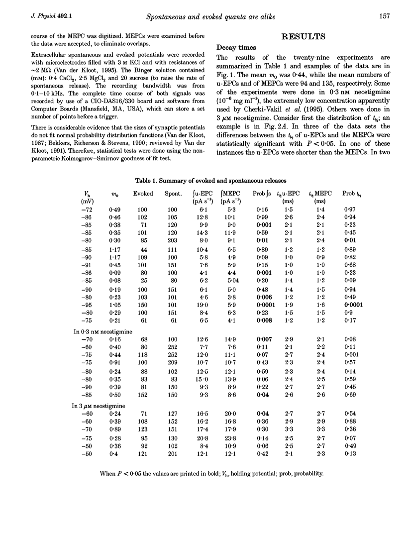
1. A recent paper concludes that the shapes of spontaneous and uniquantal-evoked signals are different. The signals were recorded with extracellular electrodes, often in the presence of neostigmine. Differences were reported between the voltage-time integrals, and between the decay times of spontaneous and evoked signals. 2. These results disagree with earlier studies using the two-electrode voltage clamp technique. 3. We recorded miniature-endplate currents (MEPCs) and uniquantal-endplate currents (u-EPCs) in a low-Mg(2+)-Ca2+ solution, sometimes with neostigmine present. Evoked quantal outputs were estimated by the method of failures, so we could reject the appropriate number of the largest evoked releases. The twenty-nine experiments showed that there were no consistent differences between the current-time integrals or half-decay times (t1/2), regardless of whether or not neostigmine was present. 4. When recording simultaneously with intracellular and extracellular electrodes, on average about 25% of the miniatures were seen in both recordings. On average, 63% of the endplate potentials were also seen in both recordings. Extracellular recording may not give the precise localization generally assumed. 5. We again conclude that quanta released by nerve stimulation and spontaneously are indistinguishable at normal frog neuromuscular junctions.
Full text
PDF


Selected References
These references are in PubMed. This may not be the complete list of references from this article.
- Barton S. B., Cohen I. S. Facilitation and impulse propagation failure at the frog neuromuscular junction. Pflugers Arch. 1982 Feb;392(4):327–334. doi: 10.1007/BF00581627. [DOI] [PubMed] [Google Scholar]
- Bekkers J. M., Richerson G. B., Stevens C. F. Origin of variability in quantal size in cultured hippocampal neurons and hippocampal slices. Proc Natl Acad Sci U S A. 1990 Jul;87(14):5359–5362. doi: 10.1073/pnas.87.14.5359. [DOI] [PMC free article] [PubMed] [Google Scholar]
- Cherki-Vakil R., Ginsburg S., Meiri H. The difference in shape of spontaneous and uniquantal evoked synaptic potentials in frog muscle. J Physiol. 1995 Feb 1;482(Pt 3):641–650. doi: 10.1113/jphysiol.1995.sp020546. [DOI] [PMC free article] [PubMed] [Google Scholar]
- Cohen I. S., van der Kloot W., Barton S. B. Bursts of miniature end-plate potentials can be released from localized regions of the frog motor nerve terminal. Brain Res. 1981 Sep 28;221(2):382–386. doi: 10.1016/0006-8993(81)90787-3. [DOI] [PubMed] [Google Scholar]
- Colméus C., Gomez S., Molgó J., Thesleff S. Discrepancies between spontaneous and evoked synaptic potentials at normal, regenerating and botulinum toxin poisoned mammalian neuromuscular junctions. Proc R Soc Lond B Biol Sci. 1982 Apr 22;215(1198):63–74. doi: 10.1098/rspb.1982.0028. [DOI] [PubMed] [Google Scholar]
- DEL CASTILLO J., KATZ B. Localization of active spots within the neuromuscular junction of the frog. J Physiol. 1956 Jun 28;132(3):630–649. doi: 10.1113/jphysiol.1956.sp005554. [DOI] [PMC free article] [PubMed] [Google Scholar]
- DEL CASTILLO J., KATZ B. Quantal components of the end-plate potential. J Physiol. 1954 Jun 28;124(3):560–573. doi: 10.1113/jphysiol.1954.sp005129. [DOI] [PMC free article] [PubMed] [Google Scholar]
- ELMQVIST D., QUASTEL D. M. PRESYNAPTIC ACTION OF HEMICHOLINIUM AT THE NEUROMUSCULAR JUNCTION. J Physiol. 1965 Apr;177:463–482. doi: 10.1113/jphysiol.1965.sp007605. [DOI] [PMC free article] [PubMed] [Google Scholar]
- Lederer W. J., Spindler A. J., Eisner D. A. Thick slurry bevelling: a new technique for bevelling extremely fine microelectrodes and micropipettes. Pflugers Arch. 1979 Sep;381(3):287–288. doi: 10.1007/BF00583261. [DOI] [PubMed] [Google Scholar]
- Molgó J., Thesleff S. 4-aminoquinoline-induced 'giant' miniature endplate potentials at mammalian neuromuscular junctions. Proc R Soc Lond B Biol Sci. 1982 Jan 22;214(1195):229–244. doi: 10.1098/rspb.1982.0006. [DOI] [PubMed] [Google Scholar]
- Thesleff S., Molgó J., Lundh H. Botulinum toxin and 4-aminoquinoline induce a similar abnormal type of spontaneous quantal transmitter release at the rat neuromuscular junction. Brain Res. 1983 Mar 28;264(1):89–97. doi: 10.1016/0006-8993(83)91123-x. [DOI] [PubMed] [Google Scholar]
- Van der Kloot W., Cohen I. S. Temperature effects on spontaneous and evoked quantal size at the frog neuromuscular junction. J Neurosci. 1984 Sep;4(9):2200–2203. doi: 10.1523/JNEUROSCI.04-09-02200.1984. [DOI] [PMC free article] [PubMed] [Google Scholar]
- Van der Kloot W., Molgó J. Quantal acetylcholine release at the vertebrate neuromuscular junction. Physiol Rev. 1994 Oct;74(4):899–991. doi: 10.1152/physrev.1994.74.4.899. [DOI] [PubMed] [Google Scholar]
- Van der Kloot W. Pretreatment with hypertonic solutions increases quantal size at the frog neuromuscular junction. J Neurophysiol. 1987 May;57(5):1536–1554. doi: 10.1152/jn.1987.57.5.1536. [DOI] [PubMed] [Google Scholar]
- Van der Kloot W. The rise times of miniature endplate currents suggest that acetylcholine may be released over a period of time. Biophys J. 1995 Jul;69(1):148–154. doi: 10.1016/S0006-3495(95)79884-8. [DOI] [PMC free article] [PubMed] [Google Scholar]
- Yu S. P., Van der Kloot W. Increasing quantal size at the mouse neuromuscular junction and the role of choline. J Physiol. 1991 Feb;433:677–704. doi: 10.1113/jphysiol.1991.sp018450. [DOI] [PMC free article] [PubMed] [Google Scholar]