Abstract
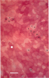
1. Using a new method of quantitative cytochemistry to determine alkaline phosphatase activity, it was possible to identify M cells and distinguish two further types of enterocytes present in mouse Peyer's patch follicle-associated epithelial tissue. 2. A quantitative description of these three cell types was provided by making direct absorbance measurements of enzyme reaction product on the luminal surface of individual follicle-associated enterocytes. M cells having a negligible alkaline phosphatase activity had a mean absorbance of 6 +/- 2 arbitrary units. The other two populations of enterocytes had overlapping but significantly different mean absorbances of 20 +/- 2 and 30 +/- 3 arbitrary units. 3. M cells accounted for 3.2 +/- 1% of the total number of cells present in the apical part of the follicle-associated epithelium. This percentage is a minimum value describing an extreme example of cells having M cell-like characteristics. The remaining epithelial cells were divided equally between those having high and low alkaline phosphatase activities in their brush-border membranes. A small percentage of cells having low alkaline phosphatase activities could perform M cell functions. 4. Results obtained suggest that reduced alkaline phosphatase activity and brush-border membrane Cl- conductance can both be used as differentiation markers to identify a large population of poorly differentiated enterocytes present in mouse follicle-associated epithelial tissue.
Full text
PDF


Images in this article
Selected References
These references are in PubMed. This may not be the complete list of references from this article.
- Cremaschi D., James P. S., Rossetti C., Smith M. W. Chloride conductance and intracellular chloride accumulation in mouse Peyer's patch enterocytes. J Physiol. 1990 Aug;427:71–80. doi: 10.1113/jphysiol.1990.sp018161. [DOI] [PMC free article] [PubMed] [Google Scholar]
- Cremaschi D., James P. S., Rossetti C., Smith M. W. Enterocyte-lymphocyte interactions in the follicle-associated epithelium of the mouse Peyer's patch. J Physiol. 1989 Jan;408:605–616. doi: 10.1113/jphysiol.1989.sp017479. [DOI] [PMC free article] [PubMed] [Google Scholar]
- Jarry A., Robaszkiewicz M., Brousse N., Potet F. Immune cells associated with M cells in the follicle-associated epithelium of Peyer's patches in the rat. An electron- and immuno-electron-microscopic study. Cell Tissue Res. 1989 Feb;255(2):293–298. doi: 10.1007/BF00224111. [DOI] [PubMed] [Google Scholar]
- Owen R. L., Bhalla D. K. Cytochemical analysis of alkaline phosphatase and esterase activities and of lectin-binding and anionic sites in rat and mouse Peyer's patch M cells. Am J Anat. 1983 Oct;168(2):199–212. doi: 10.1002/aja.1001680207. [DOI] [PubMed] [Google Scholar]
- Smith M. W., James P. S., Tivey D. R., Brown D. Automated histochemical analysis of cell populations in the intact follicle-associated epithelium of the mouse Peyer's patch. Histochem J. 1988 Aug;20(8):443–448. doi: 10.1007/BF01002430. [DOI] [PubMed] [Google Scholar]
- Smith M. W., James P. S., Tivey D. R. M cell numbers increase after transfer of SPF mice to a normal animal house environment. Am J Pathol. 1987 Sep;128(3):385–389. [PMC free article] [PubMed] [Google Scholar]
- Smith M. W., Peacock M. A. "M" cell distribution in follicle-associated epithelium of mouse Peyer's patch. Am J Anat. 1980 Oct;159(2):167–175. doi: 10.1002/aja.1001590205. [DOI] [PubMed] [Google Scholar]
- Smith M. W. Selective expression of brush border hydrolases by mouse Peyer's patch and jejunal villus enterocytes. J Cell Physiol. 1985 Aug;124(2):219–225. doi: 10.1002/jcp.1041240208. [DOI] [PubMed] [Google Scholar]


