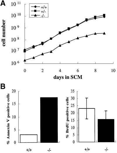
Fig. 4. Analysis of c-raf-1–/– hematopoietic cells in culture. Fetal livers were isolated on E11.5 from wild-type (+/+) and homozygous c-raf-1–/– (–/–) littermates of mixed 129BL/6 background. Cells were pre-cultured in media supporting the expansion of hematopoietic cells for 8 days to enrich for multipotent hematopoietic precursors. (A) Multipotent precursors of each genotype were seeded at a density of 2 × 10⁶ cells/ml, further cultivated and counted at daily intervals. Cells were maintained at a constant density of 2 × 10⁶ cells/ml. The plot shows the total number of cells generated per fetal liver as a function of the time in culture. (B) Percentage of annexin V-positive (left panel) or BrdU-positive (right panel) cells in asynchronous cultures (day 8) of hematopoietic fetal livers cells from wild-type (+/+, open bars) and c-raf-1–/– (–/–, closed bars) littermates. A representative experiment out of two is shown.

An official website of the United States government
Here's how you know
Official websites use .gov
A
.gov website belongs to an official
government organization in the United States.
Secure .gov websites use HTTPS
A lock (
) or https:// means you've safely
connected to the .gov website. Share sensitive
information only on official, secure websites.
