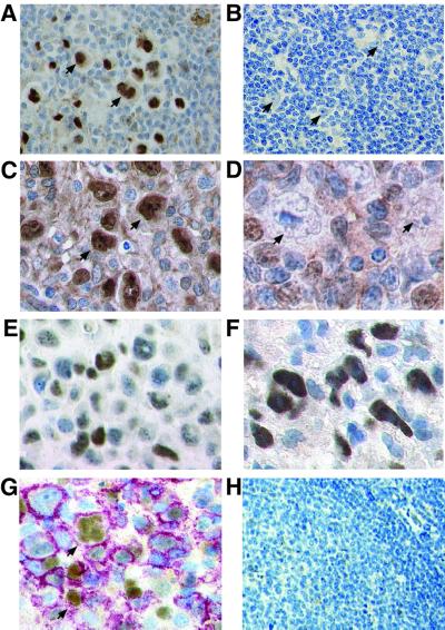
Fig. 2. High level c-Jun and JunB expression is a hallmark of malignant cells in cHD. Immunohistochemistry of representative biopsy specimen. (A) All HRS cells (arrows) in cHD, but not surrounding benign cells, reveal strong nuclear c-Jun staining. (B) Malignant cells (arrows) in LPHD do not express c-Jun in detectable amounts. (C) In all HRS cells in cHD, JunB is abundantly expressed, with mostly nuclear localization (arrows). (D) Malignant cells (arrows) in LPHD do not show JunB expression. (E) c-Jun and (F) JunB expression in ALCL. In contrast to cHD, all cells shown are tumor cells. (G) Tonsil of a patient with infectious mononucleosis analyzed by double staining for c-Jun and CD20, indicating B-cell origin. The membrane staining of CD20 appears red, nuclear staining of c-Jun, brown. B cells with HRS cell-like morphology stain positive for c-Jun (arrows). (H) Normal tonsil. No c-Jun-positive cells are detectable.

An official website of the United States government
Here's how you know
Official websites use .gov
A
.gov website belongs to an official
government organization in the United States.
Secure .gov websites use HTTPS
A lock (
) or https:// means you've safely
connected to the .gov website. Share sensitive
information only on official, secure websites.
