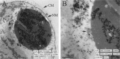
FIG. 2.
Electron micrographs of a tegument aggregate in the nucleus of a cell infected with RCΔ97. Infected cells were fixed and imaged with a Hitachi H7000 transmission electron microscope with an accelerating voltage of 75 kV. A large tegument aggregate is observed inside the nuclear membrane (NM), and the cytoplasmic membrane (CM) is shown for reference (A). A close-up of the tegument aggregate shows immature virions near the periphery of this large nuclear structure (B).

