Abstract
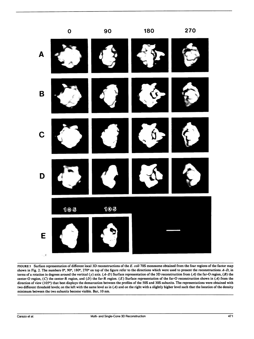
Electron microscopic techniques are among the most important tools for obtaining structural information of biological specimens. However, the three-dimensional (3D) structural analysis of asymmetrical specimens that do not form crystalline sheets has traditionally presented serious methodological obstacles to its accomplishment. One of the fundamental questions to be addressed in this type of structural study is in what way, and to what degree, does the 3D structural conformation depend on the orientation of the specimen with respect to the electron microscopic support films. As a step in studying this problem, we have analyzed the variations of the 3D structure of the Escherichia coli 70S monosome by performing four different 3D reconstructions of the 70S monosome from subsets of images in the so-called overlap range of views. These subsets were selected according to a multivariate statistical analysis performed on the total population of overlap-range specimen images. A certain amount of structural variability exists among the 3D reconstructions, although many of the main morphological characteristics, as the relative orientation between the ribosomal subunits, remain unchanged. We have also generalized the random conical reconstruction technique (Radermacher, M., T. Wagenknecht, A. Verschoor, and J. Frank. 1987. J. Microsc. 146: 113-136) to include those cases where the specimen exhibits a rocking behavior with respect to the support. The resulting Multicone Reconstruction Technique has been applied to computer-generated images as well as the E. coli 70S monosome images from part of the overlap range of views.
Full text
PDF


Images in this article
Selected References
These references are in PubMed. This may not be the complete list of references from this article.
- Arad T., Piefke J., Weinstein S., Gewitz H. S., Yonath A., Wittmann H. G. Three-dimensional image reconstruction from ordered arrays of 70S ribosomes. Biochimie. 1987 Sep;69(9):1001–1006. doi: 10.1016/0300-9084(87)90234-3. [DOI] [PubMed] [Google Scholar]
- Bretaudiere J. P., Frank J. Reconstitution of molecule images analysed by correspondence analysis: a tool for structural interpretation. J Microsc. 1986 Oct;144(Pt 1):1–14. doi: 10.1111/j.1365-2818.1986.tb04669.x. [DOI] [PubMed] [Google Scholar]
- Carazo J. M., Frank J. Three-dimensional matching of macromolecular structures obtained from electron microscopy: an application to the 70S and 50S E. coli ribosomal particles. Ultramicroscopy. 1988;25(1):13–22. doi: 10.1016/0304-3991(88)90401-9. [DOI] [PubMed] [Google Scholar]
- Carazo J. M., Wagenknecht T., Radermacher M., Mandiyan V., Boublik M., Frank J. Three-dimensional structure of 50 S Escherichia coli ribosomal subunits depleted of proteins L7/L12. J Mol Biol. 1988 May 20;201(2):393–404. doi: 10.1016/0022-2836(88)90146-5. [DOI] [PubMed] [Google Scholar]
- Frank J., Goldfarb W., Eisenberg D., Baker T. S. Reconstruction of glutamine synthetase using computer averaging. Ultramicroscopy. 1978;3(3):283–290. doi: 10.1016/s0304-3991(78)80038-2. [DOI] [PMC free article] [PubMed] [Google Scholar]
- Frank J., Verschoor A., Boublik M. Computer averaging of electron micrographs of 40S ribosomal subunits. Science. 1981 Dec 18;214(4527):1353–1355. doi: 10.1126/science.7313694. [DOI] [PubMed] [Google Scholar]
- Frank J., van Heel M. Correspondence analysis of aligned images of biological particles. J Mol Biol. 1982 Oct 15;161(1):134–137. doi: 10.1016/0022-2836(82)90282-0. [DOI] [PubMed] [Google Scholar]
- Henderson R., Unwin P. N. Three-dimensional model of purple membrane obtained by electron microscopy. Nature. 1975 Sep 4;257(5521):28–32. doi: 10.1038/257028a0. [DOI] [PubMed] [Google Scholar]
- Kellenberger E., Häner M., Wurtz M. The wrapping phenomenon in air-dried and negatively stained preparations. Ultramicroscopy. 1982;9(1-2):139–150. doi: 10.1016/0304-3991(82)90236-4. [DOI] [PubMed] [Google Scholar]
- Lake J. A. Ribosome structure determined by electron microscopy of Escherichia coli small subunits, large subunits and monomeric ribosomes. J Mol Biol. 1976 Jul 25;105(1):131–139. doi: 10.1016/0022-2836(76)90200-x. [DOI] [PubMed] [Google Scholar]
- Radermacher M., Frank J. Representation of three-dimensionally reconstructed objects in electron microscopy by surfaces of equal density. J Microsc. 1984 Oct;136(Pt 1):77–85. doi: 10.1111/j.1365-2818.1984.tb02547.x. [DOI] [PubMed] [Google Scholar]
- Radermacher M. Three-dimensional reconstruction of single particles from random and nonrandom tilt series. J Electron Microsc Tech. 1988 Aug;9(4):359–394. doi: 10.1002/jemt.1060090405. [DOI] [PubMed] [Google Scholar]
- Radermacher M., Wagenknecht T., Verschoor A., Frank J. Three-dimensional reconstruction from a single-exposure, random conical tilt series applied to the 50S ribosomal subunit of Escherichia coli. J Microsc. 1987 May;146(Pt 2):113–136. doi: 10.1111/j.1365-2818.1987.tb01333.x. [DOI] [PubMed] [Google Scholar]
- Radermacher M., Wagenknecht T., Verschoor A., Frank J. Three-dimensional reconstruction from a single-exposure, random conical tilt series applied to the 50S ribosomal subunit of Escherichia coli. J Microsc. 1987 May;146(Pt 2):113–136. doi: 10.1111/j.1365-2818.1987.tb01333.x. [DOI] [PubMed] [Google Scholar]
- Radermacher M., Wagenknecht T., Verschoor A., Frank J. Three-dimensional structure of the large ribosomal subunit from Escherichia coli. EMBO J. 1987 Apr;6(4):1107–1114. doi: 10.1002/j.1460-2075.1987.tb04865.x. [DOI] [PMC free article] [PubMed] [Google Scholar]
- Van Heel M. Angular reconstitution: a posteriori assignment of projection directions for 3D reconstruction. Ultramicroscopy. 1987;21(2):111–123. doi: 10.1016/0304-3991(87)90078-7. [DOI] [PubMed] [Google Scholar]
- Verschoor A., Frank J., Wagenknecht T., Boublik M. Computer-averaged views of the 70 S monosome from Escherichia coli. J Mol Biol. 1986 Feb 20;187(4):581–590. doi: 10.1016/0022-2836(86)90336-0. [DOI] [PubMed] [Google Scholar]
- Wittmann H. G. Architecture of prokaryotic ribosomes. Annu Rev Biochem. 1983;52:35–65. doi: 10.1146/annurev.bi.52.070183.000343. [DOI] [PubMed] [Google Scholar]
- van Heel M., Frank J. Use of multivariate statistics in analysing the images of biological macromolecules. Ultramicroscopy. 1981;6(2):187–194. doi: 10.1016/0304-3991(81)90059-0. [DOI] [PubMed] [Google Scholar]


