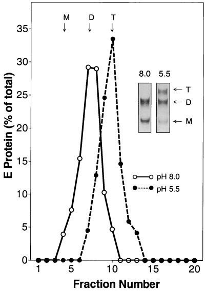
FIG. 5.
Oligomeric state of full-length E after attachment of TBE virus to liposomes. Whole TBE virions and liposomes were mixed at pH 8.0, exposed to pH 5.5, and subjected to coflotation as shown in Fig. 1. Bound virus was solubilized with 2% Triton X-100 and analyzed by sedimentation in 7 to 20% sucrose gradients containing 0.1% Triton X-100 (•). As a control, virions were incubated with liposomes at pH 8.0 and the E-protein-containing fractions were analyzed in the same way (○). The sedimentation direction is from left to right, and the positions of E monomer (M), dimer (D), and trimer (T) are indicated. (Insets) Cross-linking of the corresponding peak fractions with DMS.

