Figure 2.
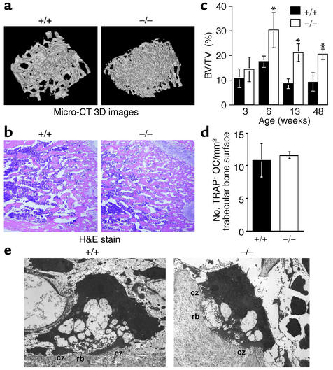
Osteopetrosis in DAP12–/– mice. (a) Micro-CT 3D images of tibial metaphyses of 40-week-old wild-type and DAP12–/– mice. DAP12–/– mice showed increased bone mass. (b) Hematoxylin and eosin staining of metaphysis sections from 6-week-old wild-type and mutant mice. (c) Percentages of trabecular bone volume per total tissue volume (BV/TV) of the femurs from wild-type (black bars) and mutant mice (white bars) at various ages. *P < 0.05. n = 3. (d) The numbers of TRAP+ multinucleated osteoclasts (OCs) in 6-week-old wild-type and DAP12–/– mice are not significantly different. (e) Transmission electron micrographs of in vivo osteoclasts from wild-type and DAP12–/– mice do not show significant differences. DAP12–/– osteoclasts displayed a ruffled border (rb), a complex membrane structure, which is usually associated with active bone matrix resorption, and a clear zone (cz) attached to the bone matrix.

