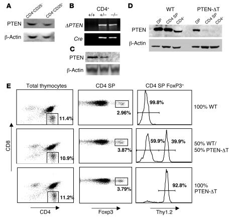
Figure 1. Thymic development of CD4+ Foxp3+ Tregs in the absence of PTEN.
(A) PTEN is expressed at equivalent levels in CD4+CD25– and CD4+CD25+ T cell subsets from normal mice. (B) Specific recombination at the Pten locus in the presence of Cre was shown by PCR amplification of an 849-bp product in genomic DNA isolated from CD4+ T cells from Cre-ve (+/+), Ptenflox/+Cre+ (+/–), and Ptenflox/floxCre+ (–/–) littermates. (C) Expression of PTEN protein in CD4+ T cells isolated as above. β-Actin was used as a loading control. (D) CD4+CD8+ double-positive (DP), CD4+ single-positive (SP), and peripheral CD4+ T cells were isolated by FACS from 3-week-old wild-type and PTEN-ΔT mice. Cells were subsequently lysed and analyzed for expression of PTEN by immunoblotting. (E) T cell–depleted bone marrow cells from wild-type (Thy1.1+) and PTEN-ΔT (Thy1.2+) mice were used to reconstitute lethally irradiated Thy1.1+ hosts. Mice were reconstituted with either 100% wild-type, 100% PTEN-ΔT, or a mixture of 50% each. Thymic regulatory subsets (CD4SP Foxp3+) were analyzed 10 weeks after reconstitution. Data shown are representative of results from 3 chimeric mice per condition.

