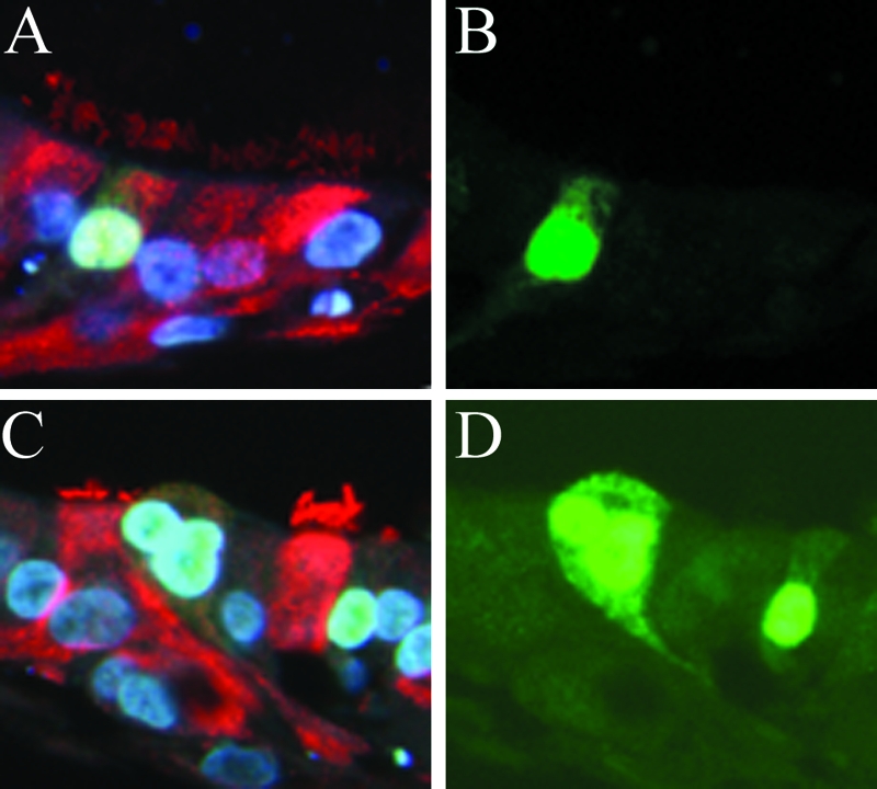
FIG. 2.

Viral proteins were detected in ciliated (A and B) and nonciliated (C and D) cells in histological cross sections of HAE infected with recent influenza A H3N2 virus strain A/England/26/99 and fixed at 6 h postinfection with 4% paraformaldehyde. Sections were costained with anti-influenza A H3N2 rabbit polyclonal antibody (green) and anti-β-tubulin IV (red) to identify ciliated cells. Cell nuclei were visualized with DAPI staining in VectorShield mounting medium. Representative fluorescent photomicrographs of viral proteins localized in the cell nuclei and cytoplasm are shown costained with β-tubulin IV and DAPI (A and C), and the same images are shown with FITC staining only (B and D).
