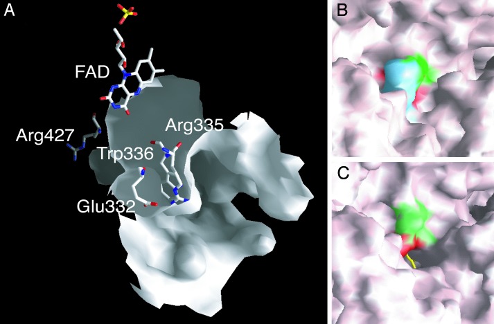
Fig. 3.
Molecular surface representation of the FAD cavity. (A) FAD and the amino acid cluster in its “open” conformation are shown in the cavity of the XO form as stick models colored by atom type. (B) The cluster in its “closed” conformation as part of the molecular surface of the XDH form. Phe-549 is shown in blue, Arg-335 is shown in green, and Trp-336 is shown in red. (C) Open conformation of the cluster as part of the molecular surface of the XO form; the accessible edge of the FAD ring is shown as a yellow stick model, and the surfaces corresponding to Arg-335 and Trp-336 are shown in green and red, respectively.

