

Abstract
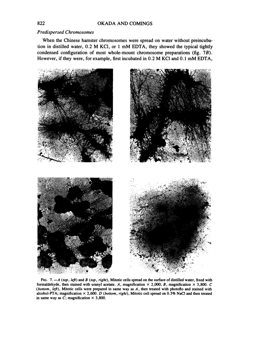
A protein chromosome scaffold structure has been proposed that acts as a structural framework for attachment of chromosomal DNA. There are several troubling aspects of this concept: (1) such structures have not been seen in many previous thin-section and whole-mount electron microscopy studies of metaphase chromosomes, while they are readily seen in leptotene and zygotene chromosomes; (2) such a structure poses problems for sister chromatid exchanges; and (3) the published photographs show a marked variation in the amount of scaffold in different whole-mount preparations. An alternative explanation is that the scaffold in whole-mount preparations represents incomplete dispersion of the high concentration of chromatin in the center of chromosomes, and when the histones are removed and the DNA dispersed, the remaining nonhistone proteins (NHPs) aggregate to form a chromosome-shaped structure. Two studies were done to determine if the scaffold is real or an artifact: (1) Chinese hamster mitotic cells and isolated chromosomes were examined using two protein stains —EDTA-regressive staining and phosphotungstic acid (PTA) stain. The EDTA-regressive stain showed ribonucleoprotein particles at the periphery of the chromosomes but nothing at the center of the chromosomes. The PTA stain showed the kinetochore plates but no central structures; and (2) isolated chromosomes were partially dispersed to decrease the high concentration of chromatin in the center of the chromosome, then treated with 4 M ammonium acetate or 2 M NaCl to dehistonize them and disperse the DNA. Under these circumstances, no chromosome scaffold was seen. We conclude that the scaffold structure is an artifact resulting from incomplete dispersion of central chromatin and aggregation of NHPs in dehistonized chromosomes.
Full text
PDF


Images in this article


Selected References
These references are in PubMed. This may not be the complete list of references from this article.
- Abuelo J. G., Moore D. E. The human chromosome. Electron microscopic observations on chromatin fiber organization. J Cell Biol. 1969 Apr;41(1):73–90. doi: 10.1083/jcb.41.1.73. [DOI] [PMC free article] [PubMed] [Google Scholar]
- Adolph K. W., Cheng S. M., Laemmli U. K. Role of nonhistone proteins in metaphase chromosome structure. Cell. 1977 Nov;12(3):805–816. doi: 10.1016/0092-8674(77)90279-3. [DOI] [PubMed] [Google Scholar]
- Adolphs K. W., Cheng S. M., Paulson J. R., Laemmli U. K. Isolation of a protein scaffold from mitotic HeLa cell chromosomes. Proc Natl Acad Sci U S A. 1977 Nov;74(11):4937–4941. doi: 10.1073/pnas.74.11.4937. [DOI] [PMC free article] [PubMed] [Google Scholar]
- Barnicot N. A., Huxley H. E. Electron microscope observations on mitotic chromosomes. Q J Microsc Sci. 1965 Sep;106(3):197–214. [PubMed] [Google Scholar]
- Berezney R., Coffey D. S. Identification of a nuclear protein matrix. Biochem Biophys Res Commun. 1974 Oct 23;60(4):1410–1417. doi: 10.1016/0006-291x(74)90355-6. [DOI] [PubMed] [Google Scholar]
- Bernhard W. A new staining procedure for electron microscopical cytology. J Ultrastruct Res. 1969 May;27(3):250–265. doi: 10.1016/s0022-5320(69)80016-x. [DOI] [PubMed] [Google Scholar]
- Chang J. P., Gibley C. W., Jr Ultrastructure of tumor cells during mitosis. Cancer Res. 1968 Mar;28(3):521–534. [PubMed] [Google Scholar]
- Comings D. E., Avelino E., Okada T. A., Wyandt H. E. The mechanism of C- and G-banding of chromosomes. Exp Cell Res. 1973 Mar 15;77(1):469–483. doi: 10.1016/0014-4827(73)90601-0. [DOI] [PubMed] [Google Scholar]
- Comings D. E., Harris D. C. Nuclear proteins. I. Electrophoretic comparison of mouse nucleoli, heterochromatin, euchromatin and contractile proteins. Exp Cell Res. 1975 Nov;96(1):161–179. doi: 10.1016/s0014-4827(75)80049-8. [DOI] [PubMed] [Google Scholar]
- Comings D. E. Mechanisms of chromosome banding and implications for chromosome structure. Annu Rev Genet. 1978;12:25–46. doi: 10.1146/annurev.ge.12.120178.000325. [DOI] [PubMed] [Google Scholar]
- Comings D. E., Okada T. A. Do half-chromatids exist? Cytogenetics. 1970;9(6):450–459. doi: 10.1159/000130114. [DOI] [PubMed] [Google Scholar]
- Comings D. E., Okada T. A. Fine structure of kinetochore in Indian muntjac. Exp Cell Res. 1971 Jul;67(1):97–110. doi: 10.1016/0014-4827(71)90625-2. [DOI] [PubMed] [Google Scholar]
- Comings D. E., Okada T. A. Some aspects of chromosome structure in eukaryotes. Cold Spring Harb Symp Quant Biol. 1974;38:145–153. doi: 10.1101/sqb.1974.038.01.018. [DOI] [PubMed] [Google Scholar]
- Comings D. E., Okada T. A. Whole mount electron microscopy of meiotic chromosomes and the synaptonmal complex. Chromosoma. 1970;30(3):269–286. doi: 10.1007/BF00321061. [DOI] [PubMed] [Google Scholar]
- Comings D. E., Okada T. A. Whole-mount electron microscopy of the centromere region of metacentric and telocentric mammalian chromosomes. Cytogenetics. 1970;9(6):436–449. doi: 10.1159/000130113. [DOI] [PubMed] [Google Scholar]
- Comings D. E. The structure and function of chromatin. Adv Hum Genet. 1972;3:237–431. doi: 10.1007/978-1-4757-4429-3_5. [DOI] [PubMed] [Google Scholar]
- Comings D. E., Wallack A. S. DNA-binding properties of nuclear matrix proteins. J Cell Sci. 1978 Dec;34:233–246. doi: 10.1242/jcs.34.1.233. [DOI] [PubMed] [Google Scholar]
- Counce S. J., Meyer G. F. Differentiation of the synaptonemal complex and the kinetochore in Locusta spermatocytes studied by whole mount electron microscopy. Chromosoma. 1973 Nov 21;44(2):231–253. doi: 10.1007/BF00329119. [DOI] [PubMed] [Google Scholar]
- Daskal Y., Mace M. L., Jr, Wray W., Busch H. Use of direct current sputtering for improved visualization of chromosome topology by scanning electron microscopy. Exp Cell Res. 1976 Jun;100(1):204–212. doi: 10.1016/0014-4827(76)90343-8. [DOI] [PubMed] [Google Scholar]
- DuPraw E. J. Evidence for a 'folded-fibre' organization in human chromosomes. Nature. 1966 Feb 5;209(5023):577–581. doi: 10.1038/209577a0. [DOI] [PubMed] [Google Scholar]
- DuPraw E. J. Macromolecular organization of nuclei and chromosomes: a folded fibre model based on whole-mount electron microscopy. Nature. 1965 Apr 24;206(982):338–343. doi: 10.1038/206338a0. [DOI] [PubMed] [Google Scholar]
- Filip D. A., Gilly C., Mouriquand C. The metaphase chromosome ultrastructure. I. Acute angle metal deposition technique as an appropriate use of shadow casting in chromosome structure investigation. Exp Cell Res. 1975 May;92(2):245–252. doi: 10.1016/0014-4827(75)90377-8. [DOI] [PubMed] [Google Scholar]
- GALL J. G. Kinetics of deoxyribonuclease action on chromosomes. Nature. 1963 Apr 6;198:36–38. doi: 10.1038/198036a0. [DOI] [PubMed] [Google Scholar]
- KRISHAN A., BUCK R. C. STRUCTURE OF THE MITOTIC SPINDLE IN L STRAIN FIBROBLASTS. J Cell Biol. 1965 Mar;24:433–444. doi: 10.1083/jcb.24.3.433. [DOI] [PMC free article] [PubMed] [Google Scholar]
- Kavenoff R., Zimm B. H. Chromosome-sized DNA molecules from Drosophila. Chromosoma. 1973;41(1):1–27. doi: 10.1007/BF00284071. [DOI] [PubMed] [Google Scholar]
- Laemmli U. K., Cheng S. M., Adolph K. W., Paulson J. R., Brown J. A., Baumbach W. R. Metaphase chromosome structure: the role of nonhistone proteins. Cold Spring Harb Symp Quant Biol. 1978;42(Pt 1):351–360. doi: 10.1101/sqb.1978.042.01.036. [DOI] [PubMed] [Google Scholar]
- Laird C. D. Chromatid structure: relationship between DNA content and nucleotide sequence diversity. Chromosoma. 1971 Mar 16;32(4):378–406. doi: 10.1007/BF00285251. [DOI] [PubMed] [Google Scholar]
- Lampert F., Lampert P. Ultrastructure of the human chromosome fiber. Humangenetik. 1970;11(1):9–17. doi: 10.1007/BF00296298. [DOI] [PubMed] [Google Scholar]
- Marsden M. P., Laemmli U. K. Metaphase chromosome structure: evidence for a radial loop model. Cell. 1979 Aug;17(4):849–858. doi: 10.1016/0092-8674(79)90325-8. [DOI] [PubMed] [Google Scholar]
- Moses M. J., Counce S. J. Electron microscopy of kinetochores in whole mount spreads of mitotic chromosomes from hela cells. J Exp Zool. 1974 Jul;189(1):115–120. doi: 10.1002/jez.1401890110. [DOI] [PubMed] [Google Scholar]
- Moses M. J. Synaptonemal complex karyotyping in spermatocytes of the Chinese hamster (Cricetulus griseus). I. Morphology of the autosomal complement in spread preparations. Chromosoma. 1977 Mar 16;60(2):99–125. doi: 10.1007/BF00288459. [DOI] [PubMed] [Google Scholar]
- Mouriquand C., Gilly C., Wolff C. Ultrastructure du chromosome: données fournies par l'observation du chromosome entier. Ann Genet. 1972 Dec;15(4):249–256. [PubMed] [Google Scholar]
- Moyne G., Garrido J. Ultrastructural evidence of mitotic perichromosomal ribonucleoproteins in hamster cells. Exp Cell Res. 1976 Mar 15;98(2):237–247. doi: 10.1016/0014-4827(76)90433-x. [DOI] [PubMed] [Google Scholar]
- Murray R. G., Murray A. S., Pizzo A. The fine structure of mitosis in rat thymic lymphocytes. J Cell Biol. 1965 Aug;26(2):601–619. doi: 10.1083/jcb.26.2.601. [DOI] [PMC free article] [PubMed] [Google Scholar]
- Okada T. A., Comings D. E. Higher order structure of chromosomes. Chromosoma. 1979 Apr 5;72(1):1–14. doi: 10.1007/BF00286426. [DOI] [PubMed] [Google Scholar]
- PORTER K. R., MACHADO R. D. Studies on the endoplasmic reticulum. IV. Its form and distribution during mitosis in cells of onion root tip. J Biophys Biochem Cytol. 1960 Feb;7:167–180. doi: 10.1083/jcb.7.1.167. [DOI] [PMC free article] [PubMed] [Google Scholar]
- Paulson J. R., Laemmli U. K. The structure of histone-depleted metaphase chromosomes. Cell. 1977 Nov;12(3):817–828. doi: 10.1016/0092-8674(77)90280-x. [DOI] [PubMed] [Google Scholar]
- ROBBINS E., GONATAS N. K. THE ULTRASTRUCTURE OF A MAMMALIAN CELL DURING THE MITOTIC CYCLE. J Cell Biol. 1964 Jun;21:429–463. doi: 10.1083/jcb.21.3.429. [DOI] [PMC free article] [PubMed] [Google Scholar]
- Rattner J. B., Krystal G., Hamkalo B. A. Selective digestion of mouse metaphase chromosomes. Chromosoma. 1978 Apr 25;66(3):259–268. doi: 10.1007/BF00330554. [DOI] [PubMed] [Google Scholar]
- Roos U. P. Light and electron microscopy of rat kangaroo cells in mitosis. I. Formation and breakdown of the mitotic apparatus. Chromosoma. 1973;40(1):43–82. doi: 10.1007/BF00319836. [DOI] [PubMed] [Google Scholar]
- Salzman N. P., Moore D. E., Mendelsohn J. Isolation and characterization of human metaphase chromosomes. Proc Natl Acad Sci U S A. 1966 Nov;56(5):1449–1456. doi: 10.1073/pnas.56.5.1449. [DOI] [PMC free article] [PubMed] [Google Scholar]
- Schwarzacher H. G., Schnedl W. Elektronenmikroskopische Untersuchungen menschlicher Metaphasen-Chromosomen. Humangenetik. 1967;4(2):153–165. doi: 10.1007/BF00291260. [DOI] [PubMed] [Google Scholar]
- Sorsa V. Condensation of chromosomes during meiotic prophase. Whole mount electron microscopy of the spermatogenesis in Omocestus. Hereditas. 1972;72(2):215–222. doi: 10.1111/j.1601-5223.1972.tb01045.x. [DOI] [PubMed] [Google Scholar]
- Sperling K., Rao P. N. The phenomenon of premature chromosome condensation: its relevance to basic and applied research. Humangenetik. 1974;23(4):235–258. doi: 10.1007/BF00272508. [DOI] [PubMed] [Google Scholar]
- Stubblefield E., Wray W. Architecture of the Chinese hamster metaphase chromosome. Chromosoma. 1971;32(3):262–294. doi: 10.1007/BF00284839. [DOI] [PubMed] [Google Scholar]
- Wolfe S. L., Hewitt G. M. The strandedness of meiotic chromosomes from Oncopeltus. J Cell Biol. 1966 Oct;31(1):31–42. doi: 10.1083/jcb.31.1.31. [DOI] [PMC free article] [PubMed] [Google Scholar]
- Wolff S. Strandedness of chromosomes. Int Rev Cytol. 1969;25:279–296. doi: 10.1016/s0074-7696(08)60205-3. [DOI] [PubMed] [Google Scholar]
- Wray W., Stubblefield E. A new method for the rapid isolation of chromosomes, mitotic apparatus, or nuclei from mammalian fibroblasts at near neutral pH. Exp Cell Res. 1970 Mar;59(3):469–478. doi: 10.1016/0014-4827(70)90656-7. [DOI] [PubMed] [Google Scholar]
- Yunis J. J., Bahr G. F. Chromatin fiber organization of human interphase and prophase chromosomes. Exp Cell Res. 1979 Aug;122(1):63–72. doi: 10.1016/0014-4827(79)90561-5. [DOI] [PubMed] [Google Scholar]