
Figure 2.
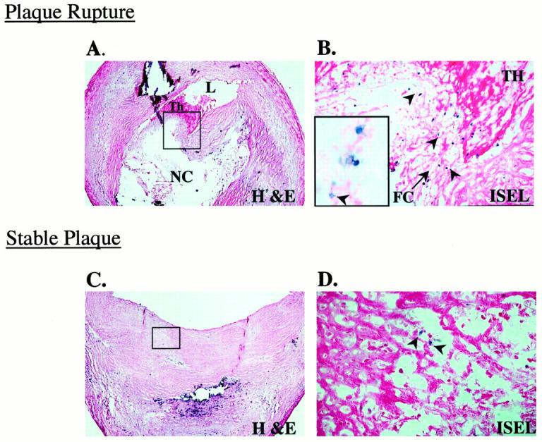
Apoptosis in culprit plaques. A: Micrograph of a cross-section of an epicardial coronary artery shows a plaque rupture with an acute luminal thrombus (Th); note the thin fibrous cap, boxed area (H&E stain; original magnification, ×30). B: Serial section of A after DNA fragmentation staining by ISEL (see Materials and Methods). Numerous apoptotic cells (blue nuclear staining, arrowheads) are identified at the plaque rupture site (eosin counterstain; original magnification, ×150). The inset shows a high-power view illustrating the nuclear detail; note the fragmented nucleus (arrowhead; original magnification, ×1000). C: Micrograph of a cross-section of an epicardial coronary artery shows an eccentric stable plaque. The lesion is characterized by dense fibrous cap, boxed area, overlying a calcified region (H&E; original magnification, ×30). D: Serial section of C after DNA fragmentation staining, there is a paucity of apoptotic cells (arrowheads) relative to rupture site in B (eosin counterstain; original magnification, ×150). Abbreviations: L, lumen; NC, necrotic core.