Figure 4.
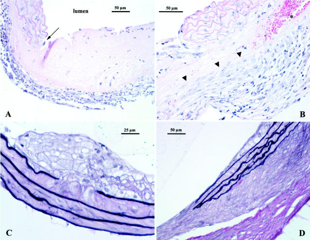
A: Day 10 lesion. Inflammatory cell infiltration of the adventitia is prominent. There is also an abrupt, focal, full-thickness necrosis of the arterial wall (arrow). This lesion appears as an acellular, pale, eosinophilic area. B: Day 20 lesion, again there is an abrupt lesion involving destruction of the vascular media. Note that activated monocytes in tissue are infiltrating into the lesion from the adventitial side (arrowheads). An area of acute hemorrhage is seen at asterisk. H&E stain (both A and B). C: Small atheroma containing foamy macrophages. Note that there is destruction of the top layer of the elastic laminae directly below the lesion. D: Extensive fibrosis in the adventitia. These changes are seen around areas where there has been destruction of the vascular media as well as beneath areas with intact elastic laminae. Note that in both C and D smooth muscle cells in the media are hypertrophied (elastin van Gieson stain, tissues from day 30).

