
Fig. 2. Immunoblot Analysis of Rat Tissue Lysates Probed with Serum from an Individual with a Presumptive Diagnosis of PANDAS.
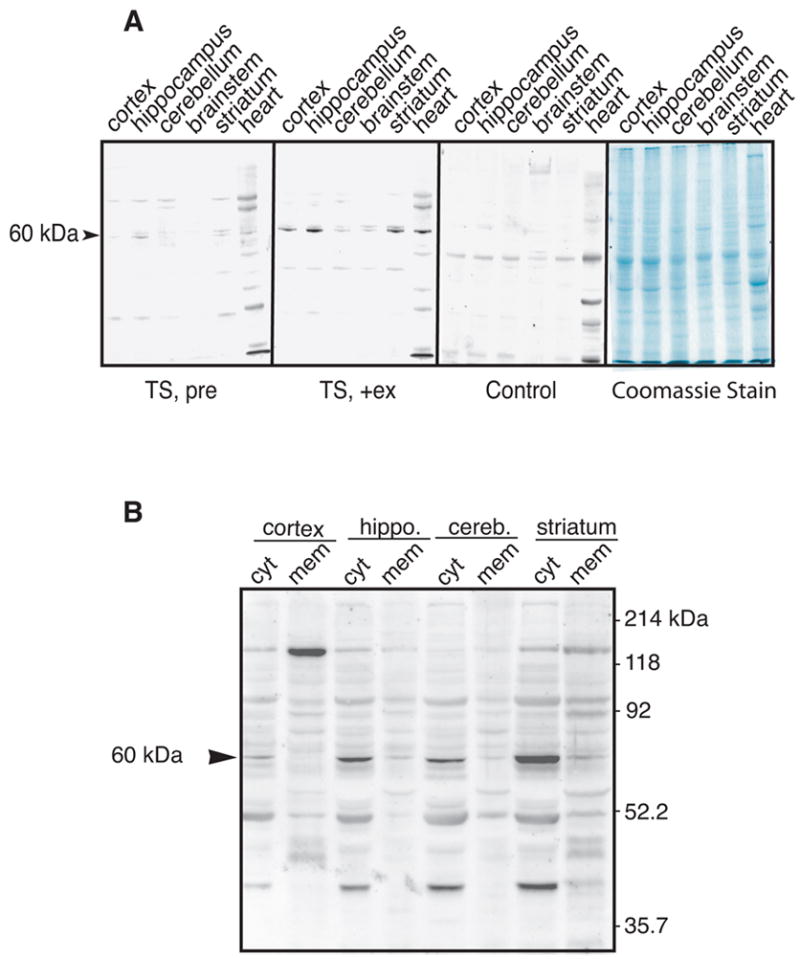
(A) The indicated tissue lysates (100 μg/lane) were blotted for reactivity to antibodies in the sera of a Tourette Syndrome (TS) patient prior to (TS, pre) and during an exacerbation of symptoms (TS,+ex). The middle right panel shows the same tissue blotted with sera from an age-matched control. The right panel shows the same tissues stained with Coomaisse blue. (B) Immunoblot of lysates (100 μg/lane) from the indicated tissue samples fractionated into cytosolic (cyt) or membrane-associated compartments (mem). The positions of the 60 kDa protein and size standards are indicated.