
Fig. 6.
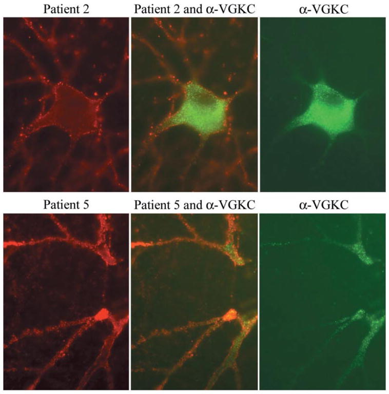
Double immunolabelling of hippocampal neuronal cultures with patients’ sera and VGKC antibodies. Using rat hippocampal neurons, the antibodies from patients 2 and 5 (red, left panels) produce widespread labelling of the surface of neurons and dendrites without co-localization (middle panels) with VGKC antibodies (green, right panels). Note that in cultured rat hippocampal neurons, the VGKC antibodies predominantly label the proximal aspect of the neuronal processes and cytoplasm. Sera of 10 other patients with radioimmunoassay-positive VGKC, and monoclonal and polyclonal antibodies to Kv1.2 produced identical reactivity to the VGKC serum used here (data not shown). All panels ×800 (oil).