
Fig. 7.
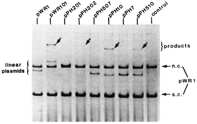
The efficiency of different att sites as linear recombination partners. The in vitro recombination reactions and gel electrophoresis were performed as described in Fig. 2 legend. Each reaction mixture contained supercoiled pWR1 (1.5μg) and linear recipient DNA in a three to one molar ratio. The linear plasmids were generated by PstI digestion of pPH7, 10, 507, 510 or by EcoRI digestion of pWR1, pWR101, pPH201 and pPH202. The origin and boundaries of the restriction fragments carried by each plasmid are described in Fig. 3 legend. Positions are indicated on the electropherogram for DNA containing different ‘recipient’ att sites (linear plasmid), the ‘donor’ circular plasmid DNA (pWR1) containing the phage att site and migrating as supercoils (s.c.) or nicked circles (n.c.) and the linear product of recombination (arrows).