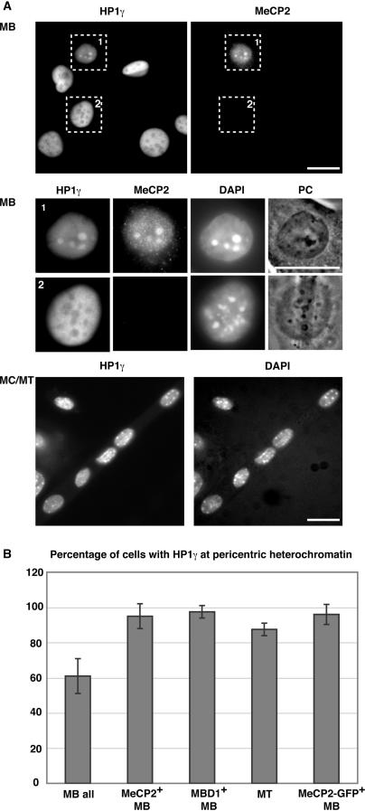
Figure 2.
Pericentric heterochromatin association of HP1γ increases during differentiation and correlates with the presence of MeCP2 and MBD1 proteins. (A) Cells were stained with HP1γ and MeCP2-specific antibodies and DNA counterstained with DAPI, highlighting the chromocenters. In the upper panels, overview images and below them representative magnified MB cells are shown, of which only the MeCP2 positive cell has HP1γ accumulated at chromocenters. The lower panels show an overview of a differentiated culture, with most nuclei having HP1γ at chromocenters. Scale bar: 20 µm. (B) Percentage of cells with HP1γ at pericentric heterochromatin and correlation with MeCP2 and MBD1 proteins. Error bars indicate SD.

