Figure 2.
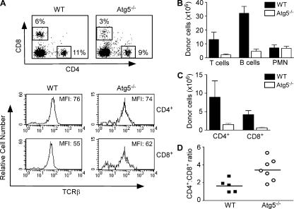
Defective peripheral lymphocyte compartment in Atg5−/− chimeras. (A) FACS profiles of donor-derived CD4+ and CD8+ T cells in the spleens of Atg5−/− and wild-type chimeric mice 6–10 wk after reconstitution. All cells are pregated on CD45.2+ donor-derived cells. (B) The numbers of T cells, B cells, and neutrophils in the spleen of Atg5−/− and wild-type chimeras. The numbers are derived from multiplying the percentage of CD4+ and CD8+ T cells, B220+, or Gr-1+CD11b+ CD45.2+ cells by the total numbers of splenocytes. p values for comparison between Atg5−/− and wild-type chimeras are: T cells, P = 0.0267; B cells, P = 0.0001; PMN, P = 0.8785. (C) The numbers of CD4+ and CD8+ T lymphocytes in the spleen of Atg5−/− and wild-type chimeras. CD4+ T cells, P = 0.0678; CD8+ T cells, P = 0.002. (D) The CD4+ to CD8+ T cell ratio in Atg5−/− and wild-type chimeras. Shown are data from individual mice. P = 0.0163. All p values are derived from unpaired, two-tailed Student's t test.

