
Figure 4.
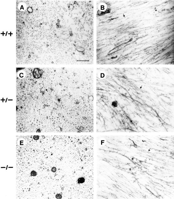
Electron microscopy of ventral root axons. Transverse sections (A, C, E) and longitudinal sections (B, D, F) of myelinated axons >5 mm in diam in the internode from the L5 ventral roots of normal (A and B) and NF-H +/− (C and D) and knockout mice (E and F). The lack of NF-H did not significantly affect the structure of 10-nm neurofilaments. Note, however, a substantial increase in the number of microtubules in both NF-H +/− and −/− mice. Open arrows, microtubules; filled arrows, 10-nm neurofilaments. Bar, 0.4 μm.