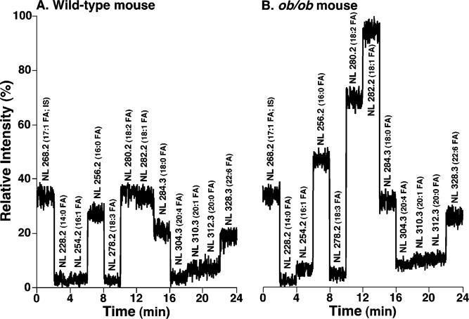
Figure 8.
Total ion current chromatogram of stepwise scanning of naturally occurring fatty acids undergoing neutral loss from TG molecular species in myocardial lipid extracts of WT and ob/ob mice at 2 months of age. Lipid extracts from WT (panel A) and ob/ob mice (panel B) were prepared by a modified Bligh and Dyer procedure as described under Materials and Methods and were analyzed in the positive-ion mode after infusion of the diluted lipid extracts in the presence of a small amount of LiOH at a flow rate of 4 μL/min. In neutral loss scanning of fatty acids, both first and third quadrupoles of a QqQ mass spectrometer were coordinately scanned with a mass difference (i.e., neutral loss) of the selected fatty acid mass while the second quadruple was used as a collision cell. The scan time between m/z 800 and m/z 960 was 1 s, and the total ion current of each scan was recorded for a 2 min segment. Neutral loss scanning of all naturally occurring fatty acids was performed sequentially as indicated with 2 min for each fatty acid using a customized program operating under Xcalibur software. Due to the facile loss of the phosphocholine headgroup, the relative contribution of fatty acyl chains from GPCho molecular species in the mass region is small in comparison to that from TG molecular species as previously described (37). Both TIC chromatograms were displayed after being normalized to the TIC of NL268.2 (i.e., 17:1 FA, the internal standard for quantitation of individual TG molecular species) as indicated.

