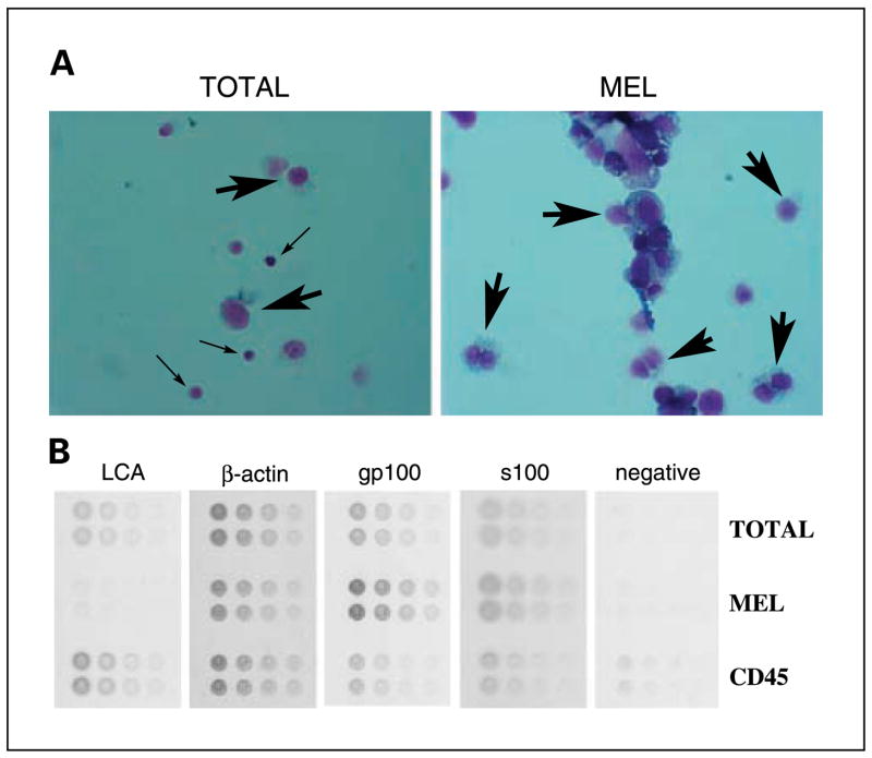
Fig. 2.
Processing of disaggregated cells from melanoma tumors by negative I-RPM. A, cytospin preparations (stained with Diffquick) reveal almost complete purity of melanoma (large arrows) in the MEL fractions and infiltrating leukocytes (small arrows) in theTOTAL fraction. B, lysates fromTOTAL, MEL, and CD45+ fractions were arrayed in minidilution format onto multiple slides.The slides were then stained with antibodies to gp100, S100, actin, and CD45 (leukocyte common antigen, LCA) or negative control.

