
Figure 10.
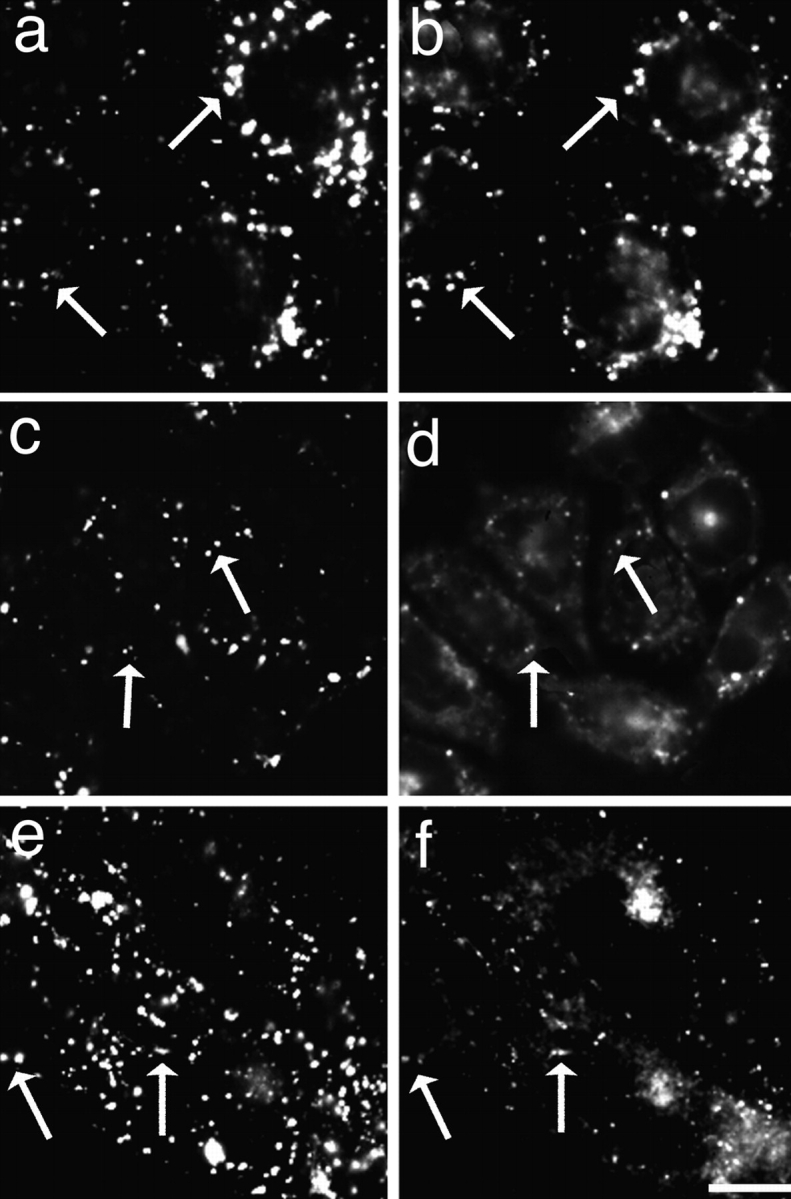
Distribution of fluorescein-dextran and the various DiI derivatives in TRVb-1 cells double labeled with 1 mg/ml fluorescein-dextran (a, c, and e) and 2 mM DiIC16(3) (b), 21 nM DiIC12(3) (d), and 75 nM FAST DiI (f), respectively. The cells were labeled for 2 min at 37°C with the DiI labeling solution, rinsed, and then incubated further at 37°C in the presence of fluorescein-dextran for 60 min. Further treatments were identical to those described in Fig. 2. Arrows indicate endosomes that contain both the fluorophores. Bar, 10 μm.