
Figure 4.
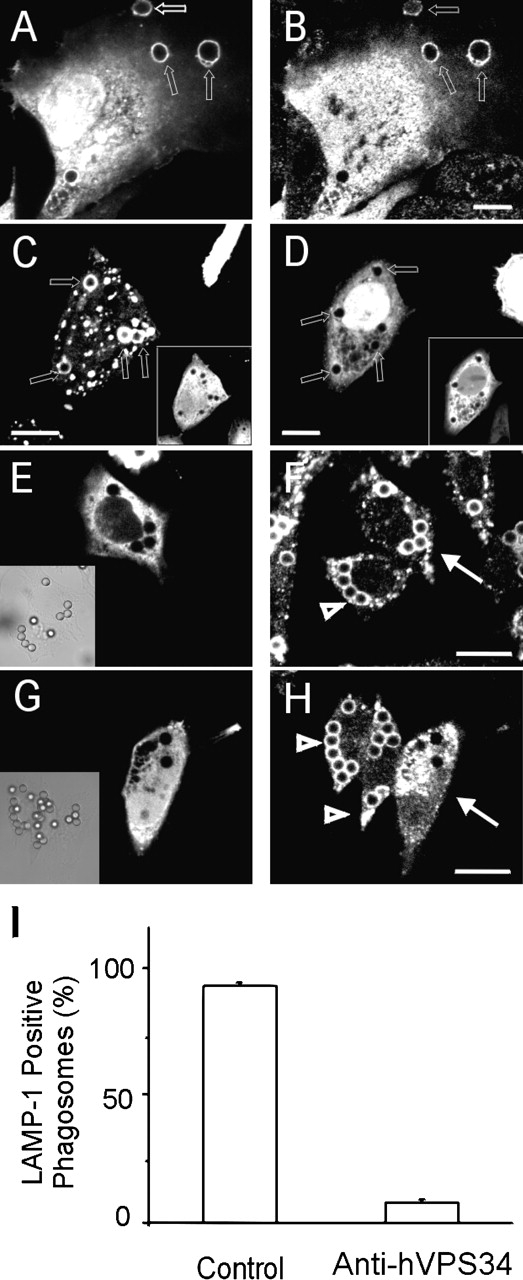
Recruitment and role of VPS34 in phago–lysosome fusion. (A and B) Distribution of 2FYVE–GFP and VPS34 during phagocytosis. COS-7 cells expressing FcγRIIA were cotransfected with 2FYVE–GFP and VPS34. After exposure to opsonized particles for 15 min, the cells were fixed and 2FYVE–GFP (A) and VPS34 (B) were visualized directly or by immunostaining, respectively. (C and D) Effect of anti-VPS34 antibodies on phagosomal distribution of 2FYVE–GFP. CHO cells transfected with 2FYVE–GFP were injected with nonimmune rabbit IgG (C) or with anti-hVPS34 antibody (D). After a 2-h period, the cells were allowed to internalize beads for 10 min. Arrows point to phagosomes. The insets identify the microinjected cells, stained with Cy3-labeled anti–rabbit IgG antibodies. (E–I) CHO cells expressing FcγRIIA were injected with either nonimmune rabbit IgG (E and F) or with anti-hVPS34 antibody (G and H). Phagocytosis of opsonized 3-μm latex beads was allowed to proceed for 20 min, followed by 50 min of maturation after removal of unbound beads, and ultimately stained for LAMP-1. (E and G) Identification of microinjected cells by staining with labeled anti-rabbit IgG. F and H: LAMP-1 staining of the cells shown in E and G, respectively. Full white arrows and open arrowheads point to injected and uninjected cells, respectively. Bars, 10 mm. (I) Quantification of phagosome acquisition of LAMP-1 in cells injected with nonimmune rabbit IgG (control) or with anti-hVPS34 antibody. Data are means ± SE of four separate experiments, each with at least 100 injected cells.