
Figure 3.
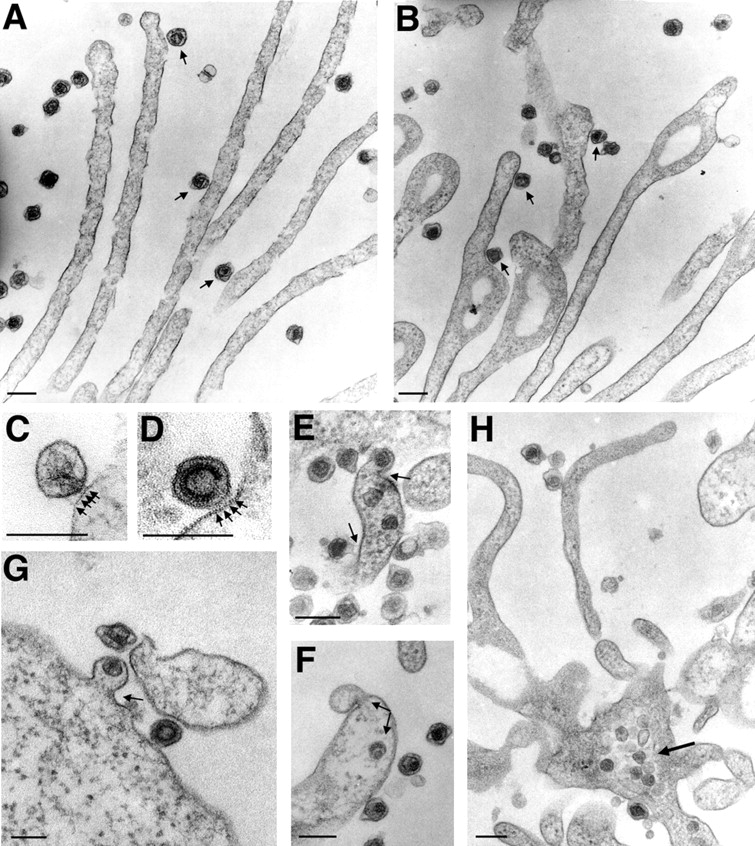
MLV fuses with the plasma membrane at the base of filopodia or at the cell body. (A and B) HEK 293 cells infected with MLV for 30 min were prepared for TEM and cut en face within 100 nm from the glass coverslip to visualize viruses and filopodia. Circular nodules as observed in B are indicative of retraction fibers (Mitchison, 1992). Viruses engaged with filopodia or retraction fibers are marked with arrows. (C and D) Two examples of frequently observed spikes (marked by arrows) between virus and membrane are shown. (E–G) Fusion sites (marked by arrows) were observed at the widening base of fibers or at the cell body. (H) Viral capsids accumulate inside the cytoplasm at the base of fibers. In all panels, size bars correspond to 200 nm.