
Figure 8.
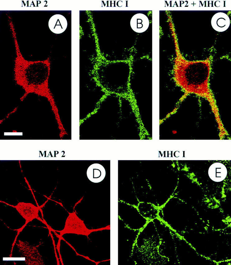
MHC class I molecules on the neuronal cell surface. Confocal laser scanning microscopy of MHC class I molecules located on the cell surface. Identification of individual neurons (pretreated with IFN-γ for 72 h) by counterlabeling the neuron-specific cytoskeleton protein MAP2. MHC class I molecules labeled red (A) and MAP2 green (B). Merger of both images from one optical section with specific labeling of MHC class I molecules on the neuronal cell surface (C). D and E show one neuron with, and another one without MHC class I cell surface labeling. Scale bar: (A–C) 10 μm; (D and E) 20 μm.