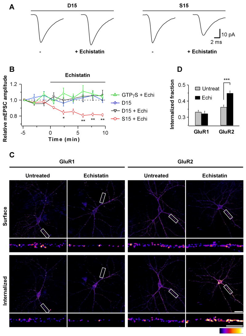
Fig 3. Integrins Regulate GluR2 Endocytosis.
(A) Neurons were loaded with D15 (left) or a scrambled S15 peptide (right) for at least 10 min prior to the baseline period. mEPSC population averages are from representative neurons during 5 min baseline (−) and the last 5 of 10 min application of echistatin (100 nM).
(B) Time course of mEPSC amplitudes from neurons loaded with GTPγS (0.5 mM; n=6), or D15 or S15 peptides (0.25–2 mM; D15, n=5; D15+echistatin, n=6; S15+echistatin, n=6). Each data point represents the average mEPSC amplitude over 100 s interval normalized to the first 100 s (*p=0.03, **p<0.01). Echistatin does not reduce mEPSC amplitudes under conditions that block endocytosis.
(C) Internalization assay for endogenous GluR1 (left) and GluR2 (right) subunits. Surface (top) vs. internalized fractions (bottom) from representative 10 min mock-(untreated) and echistatin-treated neurons (300 nM) are shown. Scale bars are 30 and 10 μm.
(D) Summary of GluR1 (n=21 images each from 4 independent cultures) and GluR2 (n=36 images each from 5 independent cultures) internalization assay. Internalized fraction indicates: (mean intensitysurface)/(mean intensitysurface + mean intensityinternalized). ***p=0.00002. Echistatin specifically increases GluR2 endocytosis.

