

Abstract
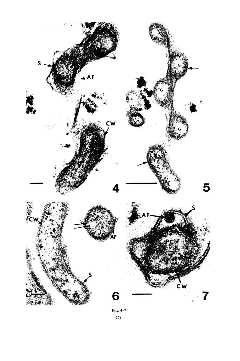
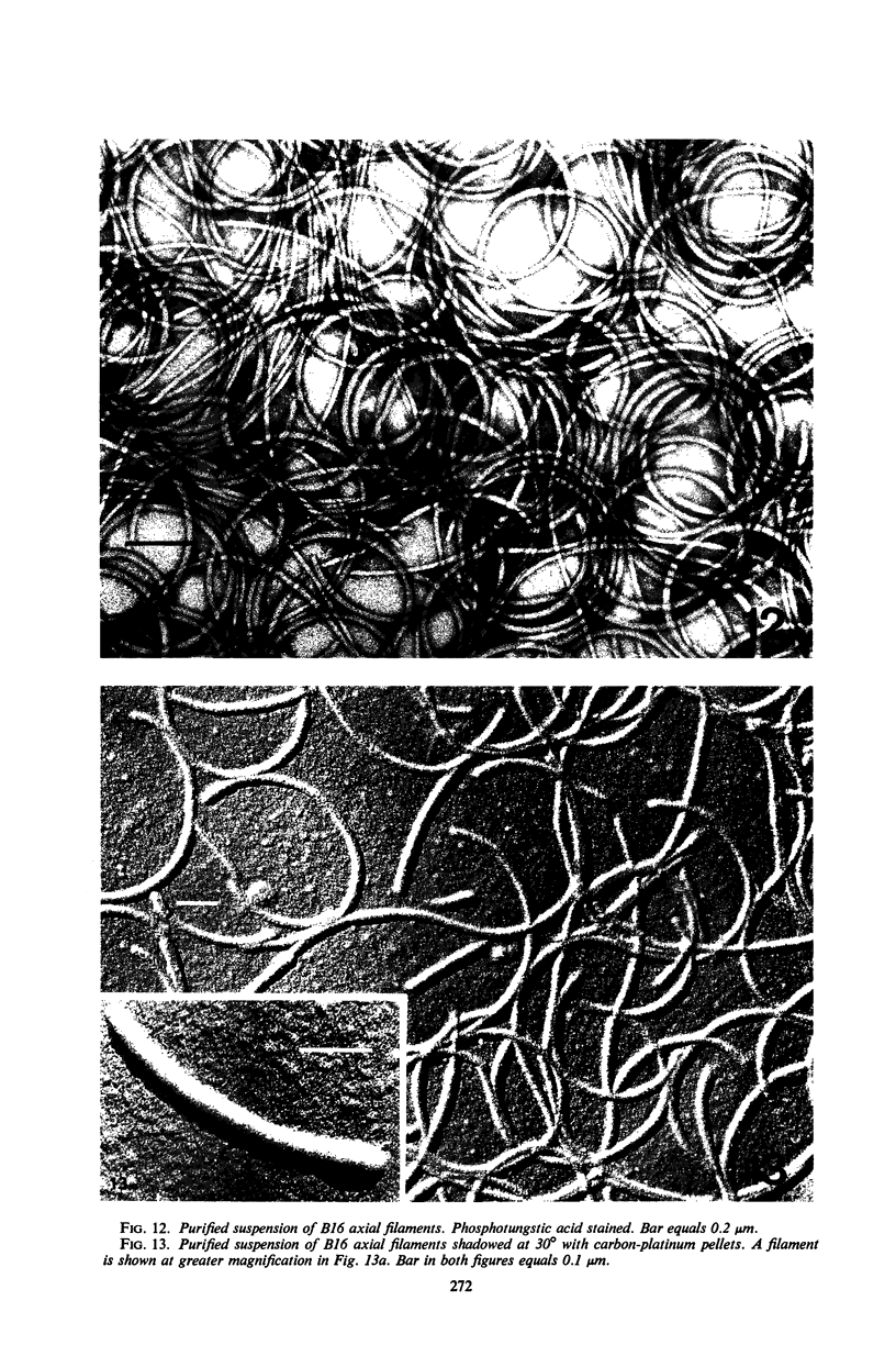
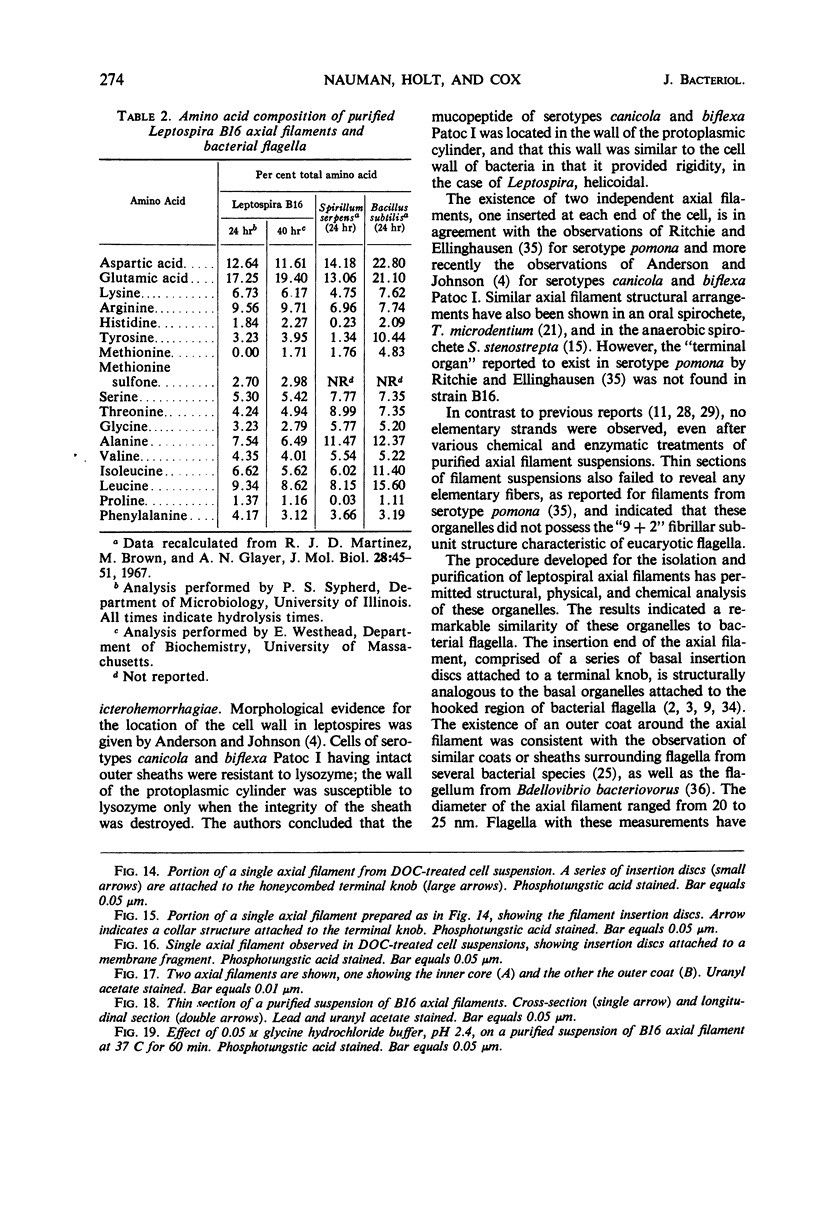
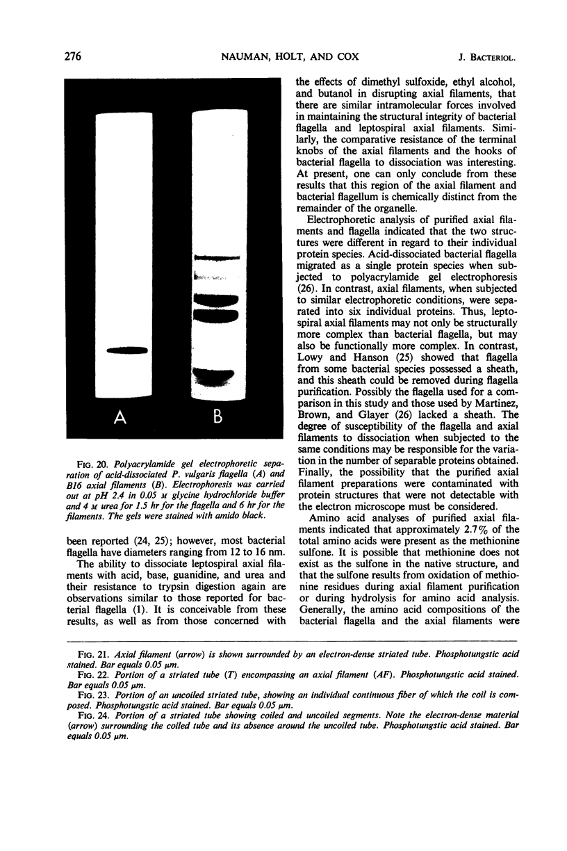
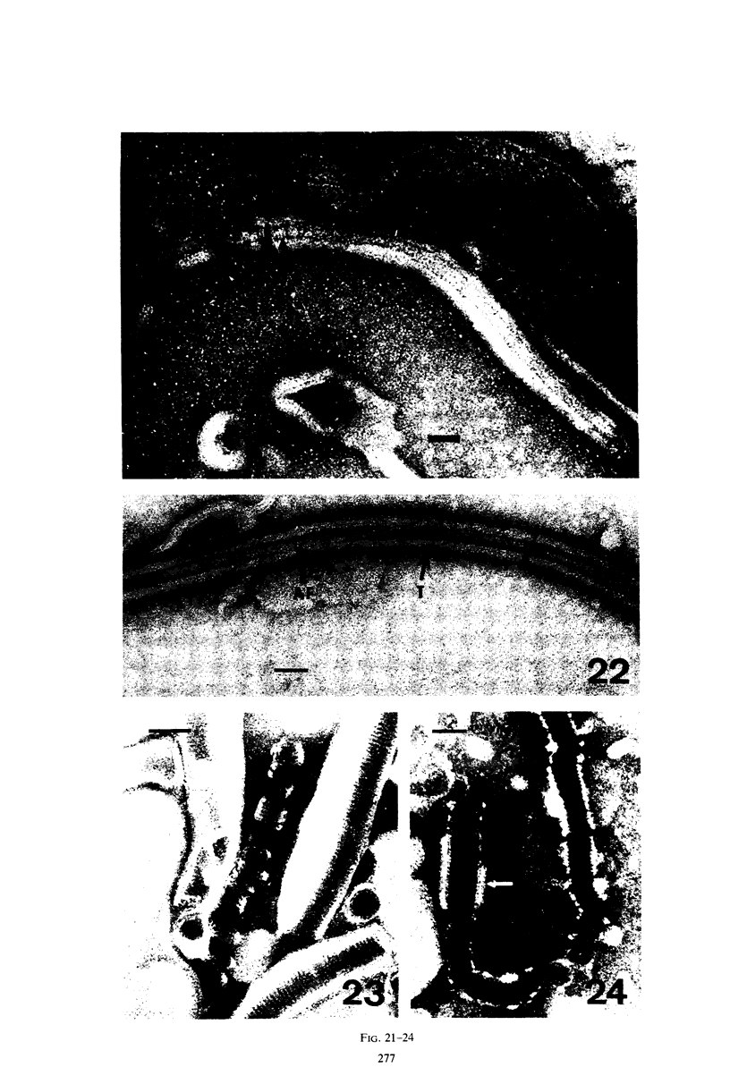
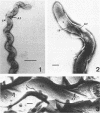
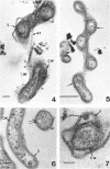
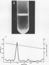
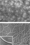
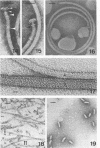
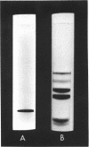
The ultrastructure of three strains of water Leptospira was studied by negative staining, thin sectioning, and freeze-etching. The cells possessed a triple-layered sheath which covered two independent axial filaments, one inserted subterminally in each end of the cell. The protoplasmic cylinder was surrounded by a triple-layered cell wall and possessed ribosomes, lamellar structures, and a typical procaryotic nuclear region. The axial filament was comprised of several component structures. An axial fibril, with a diameter of 20 to 25 nm, consisted of a solid inner core (13 to 16 nm in diameter) surrounded by a coat. A terminal knob (40 to 70 nm in length) was connected to a series of disc insertion structures at the terminal end of the axial fibril. The axial fibril was surrounded by a helical outer coat (35 to 60 nm in diameter) which was composed of a continuously coiled fiber, 3 to 4 nm in diameter, embedded in an electron-dense material. A procedure for the purification of the axial fibrils was presented and their ultrastructural, physical, and chemical properties were determined. Similarities in ultrastructural, physical, and chemical properties were noted between the axial fibrils and bacterial flagella. A schematic model of the leptospiral axial filament is presented, and a mechanism is proposed for its function as a locomotor organelle.
Full text
PDF


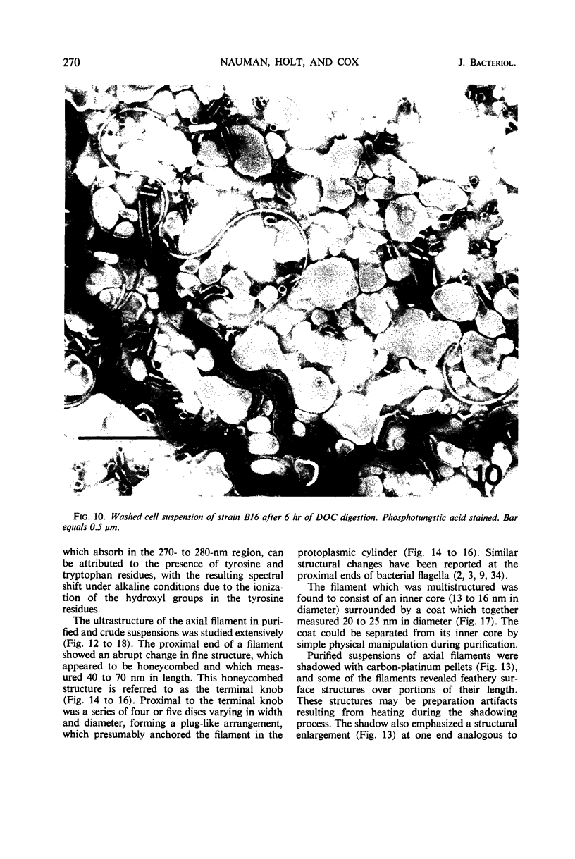


Images in this article


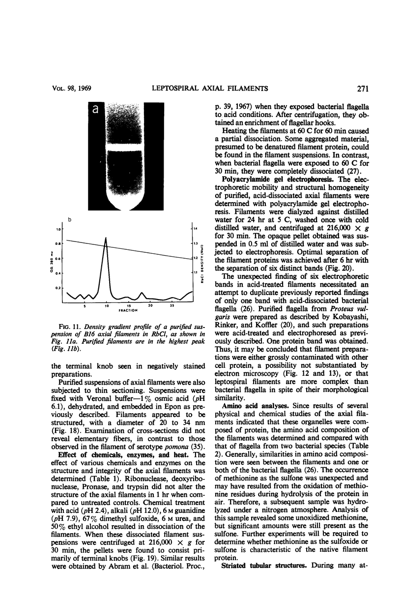
Selected References
These references are in PubMed. This may not be the complete list of references from this article.
- ABRAM D., KOFFLER H. IN VITRO FORMATION OF FLAGELLA-LIKE FILAMENTS AND OTHER STRUCTURES FROM FLAGELLIN. J Mol Biol. 1964 Jul;9:168–185. doi: 10.1016/s0022-2836(64)80098-x. [DOI] [PubMed] [Google Scholar]
- Abram D., Koffler H., Vatter A. E. Basal structure and attachment of flagella in cells of Proteus vulgaris. J Bacteriol. 1965 Nov;90(5):1337–1354. doi: 10.1128/jb.90.5.1337-1354.1965. [DOI] [PMC free article] [PubMed] [Google Scholar]
- Abram D., Vatter A. E., Koffler H. Attachment and structural features of flagella of certain bacilli. J Bacteriol. 1966 May;91(5):2045–2068. doi: 10.1128/jb.91.5.2045-2068.1966. [DOI] [PMC free article] [PubMed] [Google Scholar]
- Anderson D. L., Johnson R. C. Electron microscopy of immune disruption of leptospires: action of complement and lysozyme. J Bacteriol. 1968 Jun;95(6):2293–2309. doi: 10.1128/jb.95.6.2293-2309.1968. [DOI] [PMC free article] [PubMed] [Google Scholar]
- BABUDIERI B. [The cell structure and serology of Leptospira]. Ergeb Mikrobiol Immunitatsforsch Exp Ther. 1960;33:259–306. [PubMed] [Google Scholar]
- BRADFIELD J. R. G., CATER D. B. Electron-microscopic evidence on the structure of spirochaetes. Nature. 1952 Jun 7;169(4310):944–946. doi: 10.1038/169944a0. [DOI] [PubMed] [Google Scholar]
- Baseman J. B., Henneberry R. C., Cox C. D. Isolation and growth of Leptospira on artificial media. J Bacteriol. 1966 Mar;91(3):1374–1375. doi: 10.1128/jb.91.3.1374-1375.1966. [DOI] [PMC free article] [PubMed] [Google Scholar]
- Bladen H. A., Hampp E. G. Ultrastructure of Treponema microdentium and Borrelia vincentii. J Bacteriol. 1964 May;87(5):1180–1191. doi: 10.1128/jb.87.5.1180-1191.1964. [DOI] [PMC free article] [PubMed] [Google Scholar]
- CZEKALOWSKI J. W., EAVES G. The structure of leptospirae as revealed by electron microscopy. J Pathol Bacteriol. 1955 Jan-Apr;69(1-2):129–132. doi: 10.1002/path.1700690118. [DOI] [PubMed] [Google Scholar]
- CZEKALOWSKI J. W. Electron microscope study of Leptospira. Antonie Van Leeuwenhoek. 1963;29:29–34. doi: 10.1007/BF02046036. [DOI] [PubMed] [Google Scholar]
- Cohen-Bazire G., London J. Basal organelles of bacterial flagella. J Bacteriol. 1967 Aug;94(2):458–465. doi: 10.1128/jb.94.2.458-465.1967. [DOI] [PMC free article] [PubMed] [Google Scholar]
- Cox C. D. Studies on the isolation and growth of Leptospira from surface waters. Ann Soc Belges Med Trop Parasitol Mycol. 1966;46(2):193–202. [PubMed] [Google Scholar]
- DAVIS B. J. DISC ELECTROPHORESIS. II. METHOD AND APPLICATION TO HUMAN SERUM PROTEINS. Ann N Y Acad Sci. 1964 Dec 28;121:404–427. doi: 10.1111/j.1749-6632.1964.tb14213.x. [DOI] [PubMed] [Google Scholar]
- Henneberry R. C., Cox C. D. Antigenic analysis of water forms of Leptospira. J Bacteriol. 1968 Oct;96(4):1419–1420. doi: 10.1128/jb.96.4.1419-1420.1968. [DOI] [PMC free article] [PubMed] [Google Scholar]
- Holt S. C., Canale-Parola E. Fine structure of Spirochaeta stenostrepta, a free-living, anaerobic spirochete. J Bacteriol. 1968 Sep;96(3):822–835. doi: 10.1128/jb.96.3.822-835.1968. [DOI] [PMC free article] [PubMed] [Google Scholar]
- Holt S. C., Leadbetter E. R. Fine structure of Sporocytophaga myxococcoides. Arch Mikrobiol. 1967 Jun 21;57(3):199–213. doi: 10.1007/BF00405947. [DOI] [PubMed] [Google Scholar]
- KARNOVSKY M. J. Simple methods for "staining with lead" at high pH in electron microscopy. J Biophys Biochem Cytol. 1961 Dec;11:729–732. doi: 10.1083/jcb.11.3.729. [DOI] [PMC free article] [PubMed] [Google Scholar]
- KELLENBERGER E., RYTER A., SECHAUD J. Electron microscope study of DNA-containing plasms. II. Vegetative and mature phage DNA as compared with normal bacterial nucleoids in different physiological states. J Biophys Biochem Cytol. 1958 Nov 25;4(6):671–678. doi: 10.1083/jcb.4.6.671. [DOI] [PMC free article] [PubMed] [Google Scholar]
- KOBAYASHI T., RINKER J. N., KOFFLER H. Purification and and chemical properties of flagellin. Arch Biochem Biophys. 1959 Oct;84:342–362. doi: 10.1016/0003-9861(59)90598-3. [DOI] [PubMed] [Google Scholar]
- Kats L. N., Konstantinova N. D. Nekotorye dannye o submikroskopicheskoi strukture Leptospir. Dokl Akad Nauk SSSR. 1966 Aug 1;169(4):950–951. [PubMed] [Google Scholar]
- LISTGARTEN M. A., LOESCHE W. J., SOCRANSKY S. S. MORPHOLOGY OF TREPONEMA MICRODENTIUM AS REVEALED BY ELECTRON MICROSCOPY OF ULTRATHIN SECTIONS. J Bacteriol. 1963 Apr;85:932–939. doi: 10.1128/jb.85.4.932-939.1963. [DOI] [PMC free article] [PubMed] [Google Scholar]
- LISTGARTEN M. A., SOCRANSKY S. S. ELECTRON MICROSCOPY OF AXIAL FIBRILS, OUTER ENVELOPE, AND CELL DIVISION OF CERTAIN ORAL SPIROCHETES. J Bacteriol. 1964 Oct;88:1087–1103. doi: 10.1128/jb.88.4.1087-1103.1964. [DOI] [PMC free article] [PubMed] [Google Scholar]
- LOWRY O. H., ROSEBROUGH N. J., FARR A. L., RANDALL R. J. Protein measurement with the Folin phenol reagent. J Biol Chem. 1951 Nov;193(1):265–275. [PubMed] [Google Scholar]
- LOWY J., HANSON J. ELECTRON MICROSCOPE STUDIES OF BACTERIAL FLAGELLA. J Mol Biol. 1965 Feb;11:293–313. doi: 10.1016/s0022-2836(65)80059-6. [DOI] [PubMed] [Google Scholar]
- Lowy J. Structure of the proximal ends of bacterial flagella. J Mol Biol. 1965 Nov;14(1):297–299. doi: 10.1016/s0022-2836(65)80251-0. [DOI] [PubMed] [Google Scholar]
- MARTINEZ R. J., ROSENBERG E. THERMAL TRANSITION OF SPIRILLUM SERPENS FLAGELLA. J Mol Biol. 1964 May;8:702–707. doi: 10.1016/s0022-2836(64)80119-4. [DOI] [PubMed] [Google Scholar]
- MOLBERT E. Elektronenmikroskopische Untersuchungen zur Morphologie von Leptospiren. Z Hyg Infektionskr. 1955;141(1):82–90. [PubMed] [Google Scholar]
- MOOR H., MUHLETHALER K., WALDNER H., FREY-WYSSLING A. A new freezing-ultramicrotome. J Biophys Biochem Cytol. 1961 May;10:1–13. doi: 10.1083/jcb.10.1.1. [DOI] [PMC free article] [PubMed] [Google Scholar]
- Martinez R. J., Brown D. M., Glazer A. N. The formation of bacterial flagella. 3. Characterization of the subunits of the flagella of Bacillus subtilis and Spirillum serpens. J Mol Biol. 1967 Aug 28;28(1):45–51. doi: 10.1016/s0022-2836(67)80076-7. [DOI] [PubMed] [Google Scholar]
- Miller N. G., Wilson R. B. IN VIVO AND IN VITRO OBSERVATIONS OF LEPTOSPIRA POMONA BY ELECTRON MICROSCOPY. J Bacteriol. 1962 Sep;84(3):569–576. doi: 10.1128/jb.84.3.569-576.1962. [DOI] [PMC free article] [PubMed] [Google Scholar]
- Pillot J., Ryter A. Structure des spirochètes. 1. Etude des generes Treponema, Borrelia et Leptospira au microscope electronique. Ann Inst Pasteur (Paris) 1965 Jun;108(6):791–804. [PubMed] [Google Scholar]
- RITCHIE A. E., ELLINGHAUSEN H. C. ELECTRON MICROSCOPY OF LEPTOSPIRES. I. ANATOMICAL FEATURES OF LEPTOSPIRA POMONA. J Bacteriol. 1965 Jan;89:223–233. doi: 10.1128/jb.89.1.223-233.1965. [DOI] [PMC free article] [PubMed] [Google Scholar]
- Remsen C. C., Watson S. W., Waterbury J. B., Trüper H. G. Fine structure of Ectothiorhodospira mobilis Pelsh. J Bacteriol. 1968 Jun;95(6):2374–2392. doi: 10.1128/jb.95.6.2374-2392.1968. [DOI] [PMC free article] [PubMed] [Google Scholar]
- Ryter A., Pillot J. Structure des spirochètes. II. Etude du genre Cristispira au microscope optique et au microscope électronique. Ann Inst Pasteur (Paris) 1965 Oct;109(4):552–562. [PubMed] [Google Scholar]
- STALHEIM O. H., WILSON J. B. CULTIVATION OF LEPTOSPIRAE. II. GROWTH AND LYSIS IN SYNTHETIC MEDIUM. J Bacteriol. 1964 Jul;88:55–59. doi: 10.1128/jb.88.1.55-59.1964. [DOI] [PMC free article] [PubMed] [Google Scholar]
- Seidler R. J., Starr M. P. Structure of the flagellum of Bdellovibrio bacteriovorus. J Bacteriol. 1968 May;95(5):1952–1955. doi: 10.1128/jb.95.5.1952-1955.1968. [DOI] [PMC free article] [PubMed] [Google Scholar]
- TAKEYA K., MORI R., TODA T. Studies on the structure of Leptospira as revealed by the electron microscope. Jpn J Microbiol. 1957 Apr;1(2):99–104. doi: 10.1111/j.1348-0421.1957.tb00014.x. [DOI] [PubMed] [Google Scholar]
- Yanagawa R., Faine S. Morphological and serological analysis of leptospiral structure. Nature. 1966 Aug 20;211(5051):823–826. doi: 10.1038/211823a0. [DOI] [PubMed] [Google Scholar]