Fig. 5.
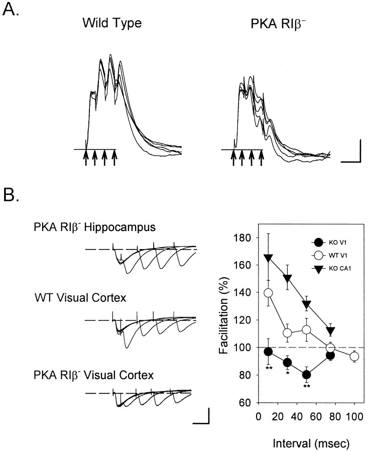
Disrupted net depolarization and paired-pulse facilitation in the visual cortex of PKA RIβ− mice.A, Whereas TBS produces a prolonged depolarization in wild-type pyramidal cells, a decrementing response is observed in the knockout cells (n = 10 of 10 cells). Whole-cell current-clamp responses to the first bursts in five episodes of TBS to layer IV are shown superimposed. Arrows indicate the four stimulus pulses delivered at 10 msec intervals. Calibration: 5 mV, 20 msec. B, Paired-pulse facilitation (PPF) is perturbed in RIβ− visual cortex but not in the hippocampus. Pairs of stimuli to layer IV elicit a prominent PPF only at 10 msec interpulse intervals in WT supragranular pyramidal cells voltage-clamped to −70 mV. In contrast, mutant V1 exhibits no PPF along this pathway, whereas it is pronounced at all intervals tested in RIβ− CA1 (n = 8 cells each; **p < 0.01, * p < 0.05;t test WT vs RIβ− cortex). Calibration: 40 pA, 20 msec.

