
Figure 1.
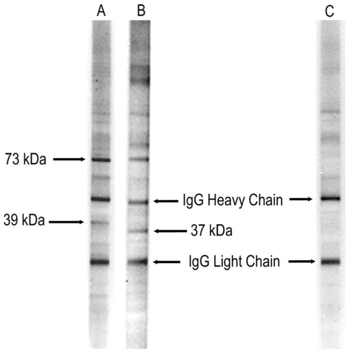
Representative lanes from western blot analysis of human fetal brain probed with serum from mothers of children with autism and those whose children are typically developing. Lane A represents a mother of a child with autism with the early onset phenotype with the 39-kDa:73-kDa band pattern. Lane B represents a mother of a child with regressive autism and the 37-kDa:73-kDa band pattern. Lane C represents a mother of a typically developing control child with no reactivity to fetal brain. The IgG heavy and light chains present in the fetal brain preparation are recognized by the secondary control and used as a reference marker.