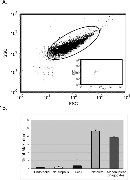
Figure 1. Analysis of the origin of peripheral blood microvesicles.
Peripheral blood microvesicles from healthy donors (n = 10) were analyzed by flow cytometry. (A) The size of the microvesicles was determined by FSC vs. SSC. The gate was set according to 2 micron beads (inset). (B) To determine cell origin, microvesicles were stained for CD3, CD202b (Tie-2), CD66b, CD79a, or CD41a to determine those that originated from T-cells, endothelial cells, neutrophils, B-cells, or platelets. Mononuclear phagocyte-derived microvesicles were positive for CD14, CD206, CCR3, CCR2, or CCR5. Shown is the average % maximum of total gated events±S.E.M.

