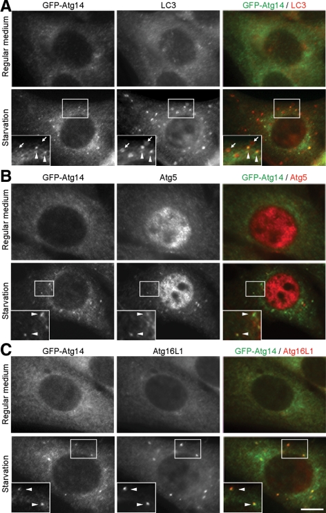
Figure 4.
Atg14 localizes on isolation membranes. NIH3T3 cells stably expressing GFP-Atg14 were cultured in regular DMEM or amino acid-free DMEM without FBS for 2 h. Then, cells were fixed, permeabilized, and subjected to immunofluorescence microscopy by using rat anti-GFP antibody and either polyclonal anti-LC3 (A), anti-Atg5 (B), or anti-Atg16L1 (C) antibody. Structures positive for both Atg14 and LC3 are indicated by arrowheads, and structures positive for LC3 but negative for Atg14 are indicated with arrows. Bar, 10 μm.

