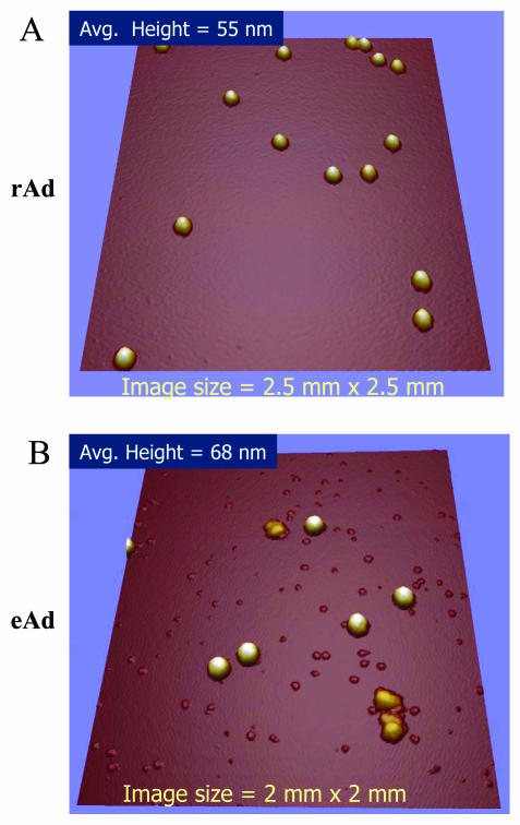
FIG. 2.
AFM of rAd and eAd. The silicon substrate surface with intact native oxide was prepared by UV cleaning, rinsing with distilled H2O (dH2O), and drying under a filtered-nitrogen stream. Approximately 15 μl of solution of 1011 Ad particles/ml was deposited on the above surface, and viruses were allowed to adsorb for 15 min. Unbound viruses were removed by washing with dH2O and then dried under a nitrogen stream. Imaging was performed in air with a ThermoMicroscopes Explorer (Veeco/TM, Sunnyvale, Calif.) with a 100-μm tripod air scanner calibrated with a NT-MDT calibration grid. Silicon tips (Nanosensors FESP; Digital Instruments, Santa Barbara, Calif.) with spring constant of ∼2 N/m and resonance frequency of ∼70 kHz were used for imaging in intermittent-contact mode. (A) rAd; (B) empty particles.

