
Figure 4.
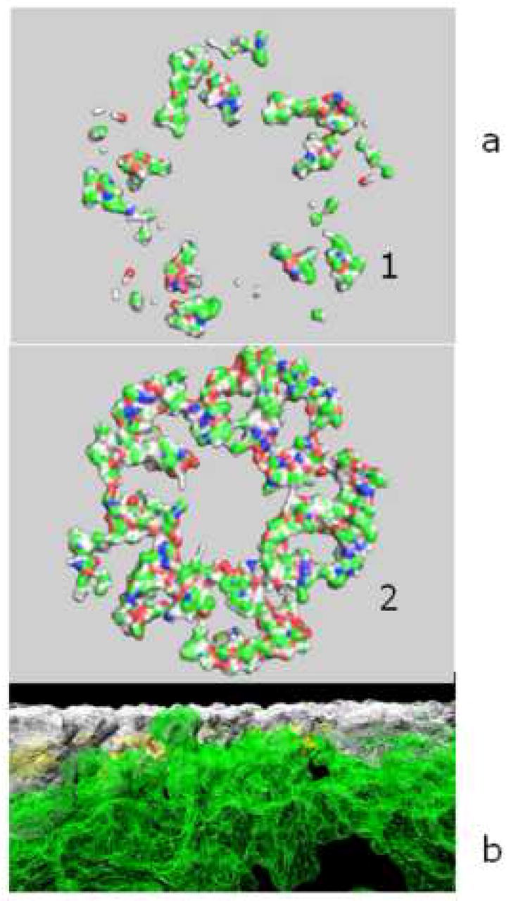
a). Portions of the synuclein-alpha (aS) pentamer that are seen behind the surface of the membrane at the beginning of MD (1) and after 0.5 ns MD (2) on the POPC membrane that are below the level of membrane surface. Pentamer is a result of docking of 4.0 ns MD aS conformers. b). Synuclein-alpha pentamer (white) embedded to the membrane (green) during 0.5 ns unrestrained MD. Pentamer is cut on the level of the top membrane atom.