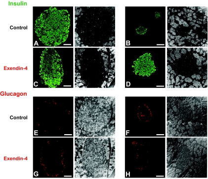
FIG. 4.
Treatment with Ex-4 improves pancreatic islet physiology in Huntington's disease mice (right panel). Immunostaining of pancreatic tissue for the β-cell–derived hormone insulin and corresponding phase contrast images. A and B: Saline-treated Huntington's disease mice had small, significantly diminished islets compared with saline-treated wild-type mice (left panel). C and D: Treatment with Ex-4 restored islet size in Huntington's disease mice but did not significantly affect islet size in wild-type mice. Immunostaining for the α-cell–derived hormone glucagon confirmed these improvements in islet physiology with Ex-4 treatment. E and G: In nondiabetic mice, glucagon-positive cells are typically arranged in a “halo” around the edge of the islet. F: In the Huntington's disease mice, the islet structure was altered, and α-cells were displaced into the center of the islet. H: This α-cell abnormality was improved with Ex-4 treatment in Huntington's disease mice. Values are means ± SE, n = 6–8 animals per group. (Please see http://dx.doi.org/10.2337/db08-0799 for a high-quality digital representation of this figure.)

