

Abstract
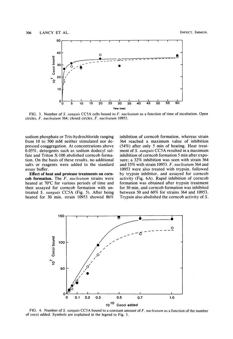
Corncob formation in dental plaque was believed to be limited to strains of Bacterionema matruchotii and Streptococcus sanguis. We observed recently that strains of Fusobacterium nucleatum also interacted with S. sanguis to form corncobs. Since the fusobacteria are among the first anaerobic filaments to colonize subgingival plaque, these interactions could serve as a connecting link between the transformation of supra- to subgingival plaque. To further characterize these interactions, quantitative in vitro studies of the kinetics of corncob formation of the fusobacteria were undertaken. These studies indicated that fewer streptococci were needed to saturate F. nucleatum strain 364 compared to strain 10953. Corncob formation with both strains was enhanced with increasing pH up to pH 8, at which point autoaggregation of the streptococci occurred. Variation in ionic strength and divalent cations had little effect on the interaction, and EDTA suppressed aggregate formation only slightly. Detergents at concentrations above 0.05% also inhibited corncob formation. Electron micrographs suggested that attachment of the cocci to the fusiforms was mediated through localized tufts of fimbriae, as they are in the Bacterionema system. However, although both trypsin and heat treatment of the streptococci inhibited corncob formation with fusobacteria, the effects were not as complete as those seen in Bacterionema species. Unlike the Bacterionema model, trypsin and heat treatment of the fusobacteria resulted in inhibition of corncob formation. These results suggest that several different receptors may be involved in corncob formation.
Full text
PDF


Images in this article

Selected References
These references are in PubMed. This may not be the complete list of references from this article.
- Appelbaum B., Golub E., Holt S. C., Rosan B. In vitro studies of dental plaque formation: adsorption of oral streptococci to hydroxyaptite. Infect Immun. 1979 Aug;25(2):717–728. doi: 10.1128/iai.25.2.717-728.1979. [DOI] [PMC free article] [PubMed] [Google Scholar]
- Bourgeau G., McBride B. C. Dextran-mediated interbacterial aggregation between dextran-synthesizing streptococci and Actinomyces viscosus. Infect Immun. 1976 Apr;13(4):1228–1234. doi: 10.1128/iai.13.4.1228-1234.1976. [DOI] [PMC free article] [PubMed] [Google Scholar]
- Carlsson J., Grahnén H., Jonsson G. Lactobacilli and streptococci in the mouth of children. Caries Res. 1975;9(5):333–339. doi: 10.1159/000260166. [DOI] [PubMed] [Google Scholar]
- Carlsson J., Grahnén H., Jonsson G., Wikner S. Establishment of Streptococcus sanguis in the mouths of infants. Arch Oral Biol. 1970 Dec;15(12):1143–1148. doi: 10.1016/0003-9969(70)90005-1. [DOI] [PubMed] [Google Scholar]
- Carlsson J. Presence of various types of non-haemolytic streptococci in dental plaque and in other sites of the oral cavity in man. Odontol Revy. 1967;18(1):55–74. [PubMed] [Google Scholar]
- Ellen R. P., Balcerzak-Raczkowski I. B. Interbacterial aggregation of Actinomyces naeslundii and dental plaque streptococci. J Periodontal Res. 1977 Jan;12(1):11–20. doi: 10.1111/j.1600-0765.1977.tb00104.x. [DOI] [PubMed] [Google Scholar]
- Gibbons R. J., Nygaard M. Interbacterial aggregation of plaque bacteria. Arch Oral Biol. 1970 Dec;15(12):1397–1400. doi: 10.1016/0003-9969(70)90031-2. [DOI] [PubMed] [Google Scholar]
- Jones S. J. A special relationship between spherical and filamentous microorganisms in mature human dental plaque. Arch Oral Biol. 1972 Mar;17(3):613–616. doi: 10.1016/0003-9969(72)90081-7. [DOI] [PubMed] [Google Scholar]
- Lancy P., Jr, Appelbaum B., Holt S. C., Rosan B. Quantitative in vitro assay for "corncob" formation. Infect Immun. 1980 Aug;29(2):663–670. doi: 10.1128/iai.29.2.663-670.1980. [DOI] [PMC free article] [PubMed] [Google Scholar]
- Listgarten M. A., Mayo H. E., Tremblay R. Development of dental plaque on epoxy resin crowns in man. A light and electron microscopic study. J Periodontol. 1975 Jan;46(1):10–26. doi: 10.1902/jop.1975.46.1.10. [DOI] [PubMed] [Google Scholar]
- Listgarten M. A., Mayo H., Amsterdam M. Ultrastructure of the attachment device between coccal and filamentous microorganisms in "corn cob" formations of dental plaque. Arch Oral Biol. 1973 May;18(5):651–656. doi: 10.1016/0003-9969(73)90105-2. [DOI] [PubMed] [Google Scholar]
- McIntire F. C., Vatter A. E., Baros J., Arnold J. Mechanism of coaggregation between Actinomyces viscosus T14V and Streptococcus sanguis 34. Infect Immun. 1978 Sep;21(3):978–988. doi: 10.1128/iai.21.3.978-988.1978. [DOI] [PMC free article] [PubMed] [Google Scholar]
- Mouton C. Association bactérienne de types différents au sein de la plaque dentaire: la formation en épi de maäis. Etude morphologique par fluorescence au microscope en lumiére ultra-violette. J Biol Buccale. 1974 Sep;2(3):207–224. [PubMed] [Google Scholar]
- Mouton C., Reynolds H. S., Genco R. J. Characterization of tufted streptococci isolated from the "corn cob" configuration of human dental plaque. Infect Immun. 1980 Jan;27(1):235–245. doi: 10.1128/iai.27.1.235-245.1980. [DOI] [PMC free article] [PubMed] [Google Scholar]
- Mouton C., Reynolds H., Genco R. J. Combined micromanipulation, culture and immunofluorescent techniques for isolation of the coccal organisms comprising the "corn cob" configuration of human dental plaque. J Biol Buccale. 1977 Dec;5(4):321–332. [PubMed] [Google Scholar]
- Newman H. N., McKay G. S. An unusual microbial configuration in human dental plaque. Microbios. 1973 Sep-Oct;8(30):117–128. [PubMed] [Google Scholar]
- Ofek I., Simpson W. A., Beachey E. H. Formation of molecular complexes between a structurally defined M protein and acylated or deacylated lipoteichoic acid of Streptococcus pyogenes. J Bacteriol. 1982 Feb;149(2):426–433. doi: 10.1128/jb.149.2.426-433.1982. [DOI] [PMC free article] [PubMed] [Google Scholar]
- Poirier T. P., Tonelli S. J., Holt S. C. Ultrastructure of gliding bacteria: scanning electron microscopy of Capnocytophaga sputigena, Capnocytophaga gingivalis, and Capnocytophaga ochracea. Infect Immun. 1979 Dec;26(3):1146–1158. doi: 10.1128/iai.26.3.1146-1158.1979. [DOI] [PMC free article] [PubMed] [Google Scholar]
- REYNOLDS E. S. The use of lead citrate at high pH as an electron-opaque stain in electron microscopy. J Cell Biol. 1963 Apr;17:208–212. doi: 10.1083/jcb.17.1.208. [DOI] [PMC free article] [PubMed] [Google Scholar]
- Ritz H. L. Microbial population shifts in developing human dental plaque. Arch Oral Biol. 1967 Dec;12(12):1561–1568. doi: 10.1016/0003-9969(67)90190-2. [DOI] [PubMed] [Google Scholar]
- Rosan B., Appelbaum B., Campbell L. K., Knox K. W., Wicken A. J. Chemostat studies of the effect of environmental control on Streptococcus sanguis adherence to hydroxyapatite. Infect Immun. 1982 Jan;35(1):64–70. doi: 10.1128/iai.35.1.64-70.1982. [DOI] [PMC free article] [PubMed] [Google Scholar]
- Slots J., Gibbons R. J. Attachment of Bacteroides melaninogenicus subsp. asaccharolyticus to oral surfaces and its possible role in colonization of the mouth and of periodontal pockets. Infect Immun. 1978 Jan;19(1):254–264. doi: 10.1128/iai.19.1.254-264.1978. [DOI] [PMC free article] [PubMed] [Google Scholar]
- Takazoe I., Matsukubo T., Katow T. Experimental formation of "corn cob" in vitro. J Dent Res. 1978 Feb;57(2):384–387. doi: 10.1177/00220345780570024101. [DOI] [PubMed] [Google Scholar]