
Figure 5. Lack of IgG in labyrinth endothelium.
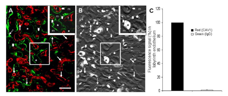
Panel A: A two-color immunfluorescence photomicrograph of the labyrinth region of a chorioallantoic placenta showing the presence of mIgG (green) and illustrating the dearth of maternal IgG in the fetal endothelium as defined by CAV1 labeling (red). The maternal blood spaces are defined by green IgG (broken arrows). Arrowheads point to trophoblast layer I & II which lack any green fluorescence signal. Asterisks are placed on the characteristically large nuclei of sinusoidal trophoblast giant cells. Bar = 30 μm. Panel B: Superimposed DAPI and DIC images are provided for orientation. Panel C: Assessing 27 mm of linear endothelium from 3 FcRn+/+ placentas (∼250 images) by 2-color immunofluorescence, we quantified the amount of IgG associated with endothelial cells (green with red). The histograms show that compared with the amount of red signal identifying endothelium (arbitrarily set at 100%), only a trace of IgG (green) colocalizes with red (≪ 1%) (P = 3 × 10⁻⁶).