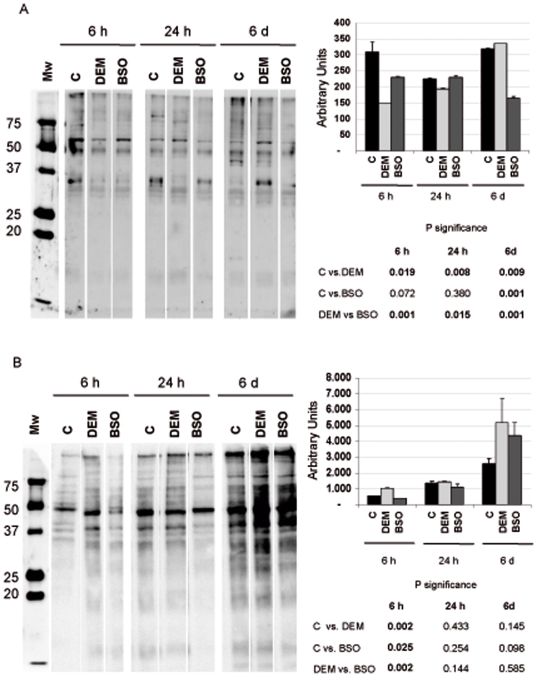
Figure 8. The pattern of overall S-glutathionylated and oxidized nuclear proteins induced by the two GSH depleting agents at 6 h, 24 h and 6 days in culture.
A.- Glutathionylated proteins: equal amount of nuclear extracts were loaded and separated by a 12% SDS gel under non-reducing conditions. S-glutathionylated proteins were detected by Western blot using anti-glutathione monoclonal antibody. B.- Oxidized proteins: equal amounts of nuclear extracts were derivatized and separated by electrophoresis in an acrilamide gel. Western blotting and subsequent immunodetection of carbonylated proteins were performed according to the protocol recommended by the manufacturer (see material and methods). Right panels shows the densitometry results for the level of glutathyolation and oxidation of nuclear proteins, respectively [Mean±SD (n = 3)].

