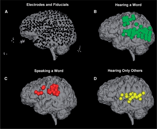
Fig. 8.
Findings from all patients are superimposed on the brain from Patient 5. (A) The locations of all of the electrodes visible over the left hemisphere (black dots) after registering them based on their fiducial points (white circles). (B) Widespread gamma activity was observed during the presentation of words (green). (C) When the patients repeated the words, activation was primarily observed over frontal and parietal cortex (red). (D) Electrodes that responded to hearing the words when presented by the computer, but not when the patient repeated the word (yellow).

