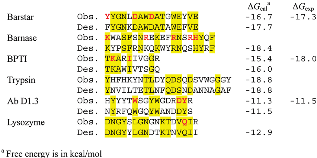
FIGURE 2.
A comparison of observed and designed interface sequences. Highlighted are the conserved residues. Hot-spot residues are shown in bold red. A residue is considered hot-spot if the binding free energy change is greater than 2.5 kcal · mol−1 upon mutation to alanine as reported for barnase–barstar (43), for BPTI (44), and for lysozyme–antibody D1.3 (47), respectively. The hot-spot residues of trypsin are not marked since the mutational information is not available.

