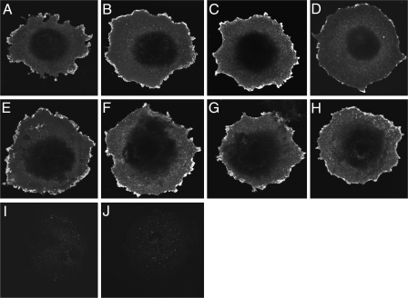
FIGURE 7.
Morphological evidence for normal cell surface expression of functionally impaired CT receptor constructs. Shown are representative examples of anti-CT receptor immunolabeling of COS-1 cells transfected with wild-type (A), Δ(1–150) (B), Δ(1–151) (C), Δ(150–154) (D), Δ(150–151) (E), Y150A (F), L151A (G), and I153A (H) CT receptors and cells transfected with the empty pcDNA3 eukaryotic expression vector (I). Shown also are COS-1 cells transfected with the wild-type CT receptor (J) but immunostained only with Alexa Fluor 488-conjugated goat anti-rabbit IgG. Images are representative of three repetitions.

