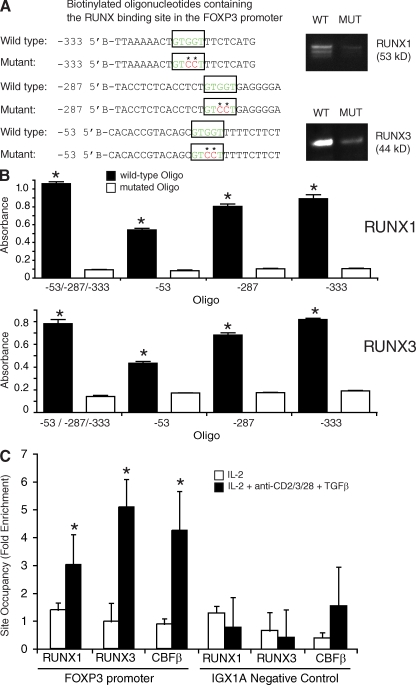
Figure 3.
Binding of RUNX1 and RUNX3 proteins to the predicted binding sites in the FOXP3 promoter. (A) Mutated and wild-type oligonucleotides are shown. The predicted RUNX binding sites are accentuated (boxed and in green letters) and stars mark mutations introduced into the binding site of the control oligonucleotides. Nuclear extracts from HEK293T cells were incubated with biotinylated oligonucleotides. The precipitated oligonucleotide–transcription factor complexes were separated by SDS-PAGE and identified by Western blotting with anti-RUNX1 and anti-RUNX3 antibodies. A mixture of all three oligonucleotides with the predicted binding sites or with the inserted mutation into the predicted sites was used. Data shown are one representative of three independent experiments with similar results. (B) Promoter enzyme immunoassay using wild-type and mutated oligonucleotides within the FOXP3 promoter. Bars show mean ± SE of three independent experiments. (C) Chromatin immunoprecipitation assay results show binding of RUNX1 and RUNX3 complexes containing CBFβ to the human FOXP3 promoter in naive CD4+ T cells that were cultured with IL-2 together with anti-CD2/3/28 and TGF-β. There was no change in site occupancy in all immunoprecipitations when IGX1A negative control primers were used. The results are normalized to input and isotype control antibody. Bars show mean ± SE of three independent experiments. Statistical differences were verified by the paired Student's t test. *, P < 0.05

