Figure 11.
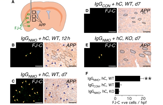
Intra-cerebral injection of IgGNMO + hC causes axonal injury within 12 h and neuronal cell death within 7 days. (A) Schematic showing needle and areas examined for FJ-C stain (green rectangles) and βAPP immunostain (black rectangles). (B–E) Representative areas stained with FJ-C (left) or immunostained with βAPP (right). Yellow arrowheads indicate FJ-C-positive cells, blue arrowheads show βAPP immunopositivity, dashed black lines demarcate degenerating white matter tracts and continuous black lines demarcate intact white matter tracts. (F) Data summary of FJ-C-positive cells/high power field at Day 7. Mean ± SEM. N = 6 (IgGNMO, hC, WT), 5 (IgGCON, hC, WT), 5 (IgGNMO, hC, KO); four high-power fields per mouse. Bar 50 µm (B–E, left), 100 µm (B–E, right). **P < 0.005 compared with IgGCON, hC, WT and with IgGNMO, hC, KO.

