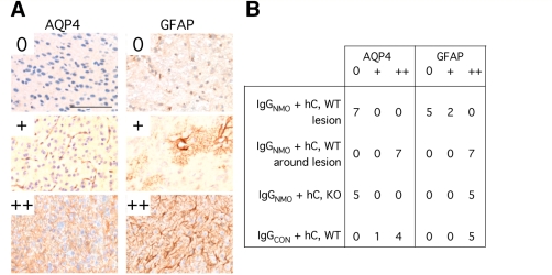
Figure 7.
Quantification of AQP4 and GFAP immunoreactivities 7 days after intra-cerebral injection of IgGNMO + hC or IgGCON + hC. (A) AQP4 expression: 0 = nil; += perivascular; ++ = parenchymal. GFAP expression: 0 = nil; + = perivascular and/or occasional reactive astrocyte; ++ = high density of reactive astrocytes. (B) Summary of AQP4 and GFAP immunoreactivities in and around lesions of WT mice injected with IgGNMO + hC, and next to the needle tracts of AQP4-null (KO) mice injected with IgGNMO + hC or WT mice injected with IgGCON + hC. Bar 100 µm (all panels).

