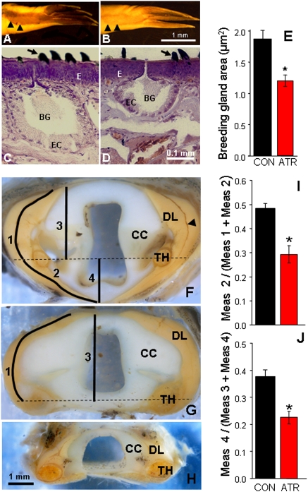
Fig. 3.
Atrazine-demasculinized male morphology as shown in the nuptial glands and the larynx. (A and B) Forearms, showing nuptial pads from control (A) and atrazine-exposed males (B). Note the reduced nuptial pads in the atrazine-exposed male (B). Black arrowheads in A and B show boundaries of nuptial pads. (C and D) Representative largest breeding gland (selected from the midpoint of the nuptial pad) from control (C) and atrazine-exposed (D) males. The area of the largest section of the largest gland was determined for each sample. Control males had significantly larger glands (E). (F–H) Transverse cross-sections through the dissected larynges of a representative sexually mature control male (F), atrazine-exposed male (G), and control female (H) X. laevis. Atrazine-exposed males had a laryngeal morphology intermediate between unexposed males and females. The dilater larynges (DL) extended well beyond the thiohyral (TH) in control males, but very little (or not at all, as in the example shown) in atrazine-exposed males. This measure was quantifiable and significantly different between controls and atrazine-exposed animals, regardless of whether the absolute length of the muscle was measured (I) or the straight-line distance (J). Black arrowhead in F indicates the slip of the dilator larynges. Horizontal dashed lines in F and G indicate the midpoint of the thiohyral. ATR, atrazine-exposed; BG, breeding gland; CC, cricoid cartilage; CON, control; E, epidermis; EC, epithelial cells. *P < 0.05; n = 14 for breeding glands, n = 11 for larynges. (Scale bar in B applies to A and B; in D applies to C and D; in H applies to F–H.)

