
Figure 3.
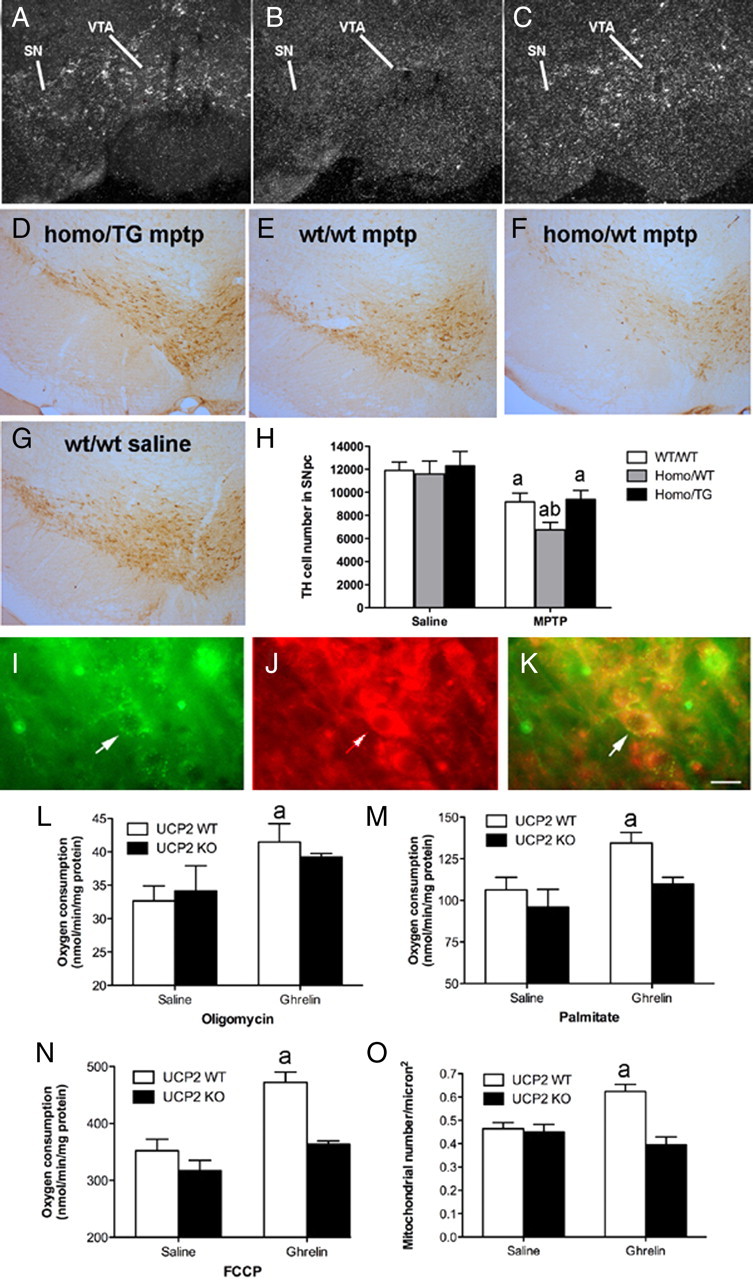
Selective reactivation of GHSR TH neurons restricts MPTP-induced DA cell loss. A–C, Representative dark-field photomicrographs of in situ hybridization histochemistry experiments performed on mouse brains using a mouse GHSR-specific riboprobe. Shown are GHSR mRNA expression within the substantia nigra (SN) and ventral tegmental area (VTA) of wild-type mice (A), the lack of GHSR transcripts within the SN and VTA of ghsr−/− mice (B), and reactivated GHSR mRNA expression within the SN and VTA of “homo/TG” mice (C). D–G, Representative low-power images of TH staining in the SNpc after MPTP insults (n = 6 all groups) in all genotypes (E–G) compared with w/w saline (D). H, Unbiased stereological quantification of total TH cell number in the SNpc of mice treated with saline or MPTP.aSignificant with respect to saline-treated control mice. bSignificant with respect to w/w MPTP-treated mice (p < 0.05). I–O, Ghrelin promotes UCP2-dependent mitochondrial respiration and proliferation. I, Immunofluorescent image showing GHSR in the SNpc. J, UCP2 in the SNpc using β galactosidase, which is the protein encoded by the lacZ reporter gene. K, Merge showing that GHSRs and UCP2 are coexpressed in SNpc neurons. Scale bar, 1 μm. L, Ghrelin increases mitochondrial respiration after the addition of oligomycin in wt but not ucp2−/− mice (10 nmol, i.p.; n = 4). M, Elevated uncoupling activity in UCP2 wt but not ucp2−/− mice after ghrelin treatment as indicated by increased mitochondrial respiration after the addition of the free fatty acid palmitate (n = 4). N, FCCP increases total respiratory capacity in UCP2 wt but not ucp2−/− mice after ghrelin injection (n = 4, each sample represents 3 pooled midbrain dissections). O, Increased mitochondrial number in SNpc DA neurons after ghrelin injection 3 h earlier (10 nmol, n = 13–15). Data are presented as the mean ± SEM. aSignificant respect to wt saline.