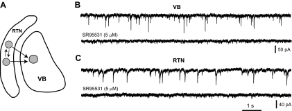
Fig. 1.
GABAergic transmission in the thalamus. A: schematic view of GABAergic circuits in the ventrobasal complex (VB) and reticular thalamic nucleus (RTN). GABAergic neurons in the RTN provide the main inhibitory input to VB neurons and are interconnected through GABAergic synapses. B and C: spontaneous inhibitory postsynaptic currents (IPSCs) recorded in a VB (B) and RTN (C) neuron from wild-type (WT) mice at P14 in the absence (top traces in B and C) and presence of the specific GABAA antagonist SR95531 (bottom traces in B and C).

